Bio 7

Exam 2: Ch 26, 27, 28

Phylogeny · prokaryotes · protists

Genus names are italicized and underlined. Blue = big idea · yellow = what she stressed · green = memory hooks · pink = be able to draw/explain and traps.

  1. 26.1 Phylogenies show evolutionary relationships
  2. 26.2 Phylogenies are inferred from morphological and molecular data
  3. 26.3 Shared characters are used to construct phylogenetic trees
  4. 26.4 An organism's evolutionary history is documented in its genome
  5. 26.5 Molecular clocks help track evolutionary time
  6. 26.6 Our understanding of the tree of life continues to change based on new data
  7. 27.1 Structural and functional adaptations contribute to prokaryotic success
  8. 27.2 Rapid reproduction, mutation, and genetic recombination promote genetic diversity in prokaryotes
  9. 27.3 Diverse nutritional and metabolic adaptations have evolved in prokaryotes
  10. 27.4 Prokaryotes have radiated into a diverse set of lineages
  11. 27.5 Prokaryotes play crucial roles in the biosphere
  12. 27.6 Prokaryotes have both beneficial and harmful impacts on humans
  13. PV Phylogenetics videos: taxonomy, homology vs analogy, and the anole lizards
  14. 28.1 Most eukaryotes are single-celled organisms
  15. 28.2 Excavates include protists with modified mitochondria and protists with unique flagella
  16. 28.3 The "SAR" clade is a highly diverse group of protists defined by DNA similarities
  17. 28.4 Red algae and green algae are the closest relatives of plants
  18. 28.5 Unikonts include protists that are closely related to fungi and animals
  19. 28.6 Protists play key roles in ecological communities
26.1

Phylogenies show evolutionary relationships

Lecture video 26.1 (opens online)
Big idea
Phylogeny is the evolutionary history of a species or group of related species, and systematics is the discipline that classifies organisms and determines their evolutionary relationships. Linnaeus (18th century) gave us binomial nomenclature and hierarchical classification, both still used. A phylogenetic tree is a hypothesis about evolutionary relationships: branch points are common ancestors, and the branching pattern (not the order of the tips) shows the order in which lineages diverged. Trees show patterns of descent, not phenotypic similarity, and do not show when species evolved or how much change occurred.

Must know

Terms

phylogeny
The evolutionary history of a species or group of related species.
systematics
The discipline that classifies organisms and determines their evolutionary relationships.
binomial nomenclature
Linnaeus's two-part scientific name for a species: genus plus specific epithet.
genus
First part of the binomial; always capitalized.
specific epithet
Second part of the binomial; unique for each species within the genus; not capitalized.
hierarchical classification
Grouping species in increasingly inclusive categories, from species up to domain.
taxon (taxa)
A named group at any level of the hierarchy.
phylogenetic tree
A branching diagram that represents a hypothesis about the evolutionary history of a group.
branch point (node)
The point where two lineages diverge from a common ancestor.
evolutionary lineage
A sequence of ancestral organisms leading to a particular descendant taxon.
sister taxa
Groups that share an immediate common ancestor that is not shared by any other group.
rooted tree
A tree that includes a branch for the most recent common ancestor of all taxa in the tree.
basal taxon
A lineage that diverges early in the history of the group, off on its own branch near the root.
polytomy
A branch point from which more than two lineages emerge; it means the order of divergence is not yet resolved. (Term assigned by the study guide request; not stated in the lecture audio.)

Examples

What she stressed in lecture
Memory hooks

Slides

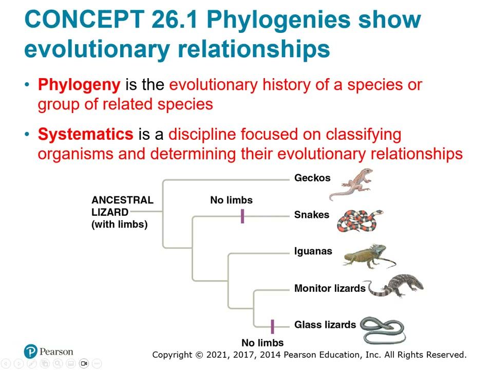
Slide 1. Limb loss in lizards: snakes vs glass lizards
No limbs evolved twice: once in snakes and once in glass lizards. Glass lizards look like snakes but are more closely related to monitor lizards, so a phylogeny shows descent, not resemblance.▶ Play from 0:07Lecture 26.1, slide 002
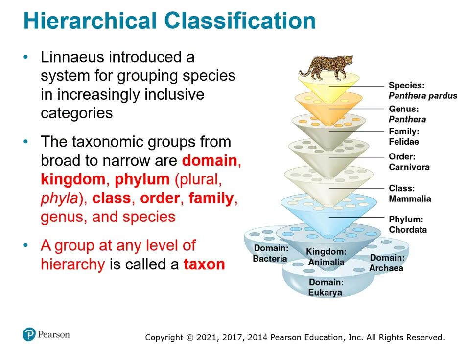
Slide 2. Hierarchical classification of the leopard
Linnaeus grouped species in increasingly inclusive categories, shown here for Panthera pardus. Know the order from broad to narrow: domain, kingdom, phylum, class, order, family, genus, species; a group at any level is a taxon.
How to draw it: Draw the nested levels for the leopard and label each: Domain Eukarya, Kingdom Animalia, Phylum Chordata, Class Mammalia, Order Carnivora, Family Felidae, Genus Panthera, Species Panthera pardus.
▶ Play from 5:40Lecture 26.1, slide 005
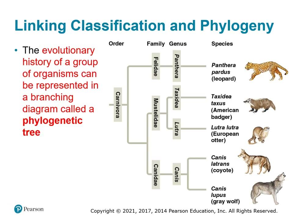
Slide 3. Linking classification and phylogeny: order Carnivora
A phylogenetic tree of Carnivora with the families Felidae, Mustelidae and Canidae and their genera. The branching matches the Linnaean groups, so Canis latrans and Canis lupus share the most recent common ancestor.▶ Play from 8:46Lecture 26.1, slide 007
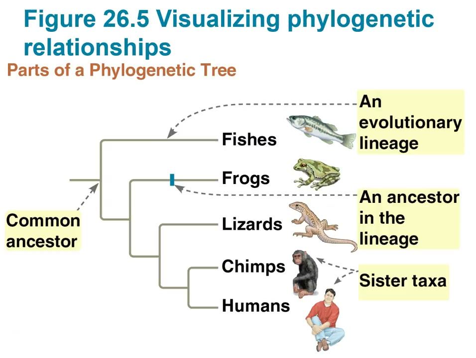
Slide 4. Parts of a phylogenetic tree
Each branch point is the divergence of two lineages from a common ancestor, and sister taxa (chimps and humans) share an ancestor not shared by any other group. The branch at the base marks the common ancestor of all taxa, which makes the tree rooted.
How to draw it: Draw a rooted tree of fishes, frogs, lizards, chimps and humans; label the common ancestor, one branch point, an evolutionary lineage, an ancestor in the lineage, and the sister taxa (chimps and humans).
▶ Play from 14:25Lecture 26.1, slide 011
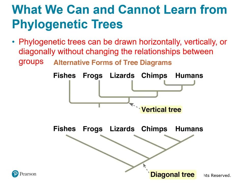
Slide 5. Vertical and diagonal forms of the same tree
The same five taxa drawn as a vertical tree and a diagonal tree. Tree orientation does not change the relationships; only the branching pattern matters.▶ Play from 16:32Lecture 26.1, slide 012
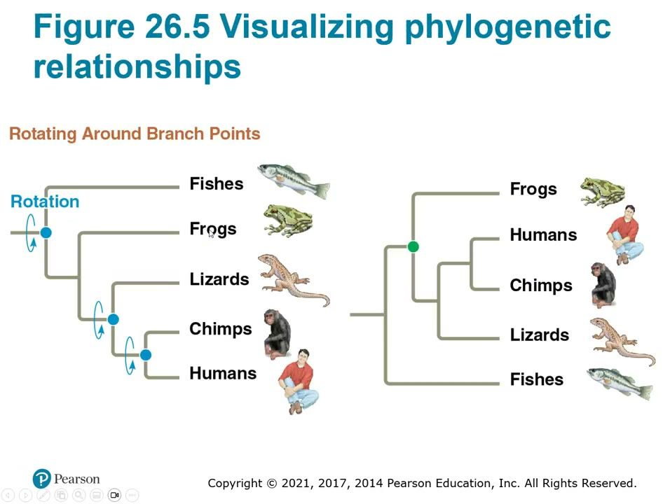
Slide 6. Rotating around branch points
Branches rotate around a branch point without changing relationships, so both trees say the same thing. The order of taxa at the tips is not significant, and a taxon did not evolve from the taxon next to it.▶ Play from 18:07Lecture 26.1, slide 016
Be able to draw / explain
My notes
26.2

Phylogenies are inferred from morphological and molecular data

Lecture video 26.2 (opens online)
Big idea
Systematists use morphology, genes (DNA and protein sequences), and biochemistry to infer phylogeny. Only features that result from common ancestry (homologies) are useful. Analogy is similarity due to convergent evolution, and it must be sorted out from homology before building a tree. The more complex the shared similarity, the more likely it reflects common ancestry.

Must know

Terms

homology
Similarity (phenotypic or genetic) due to shared ancestry.
analogy
Similarity due to convergent evolution rather than shared ancestry.
convergent evolution
Independent evolution of similar features in unrelated species exposed to similar selection pressures.
homologous structures
Structures similar because they were inherited from a common ancestor, even if they now serve different functions.
analogous structures
Structures with similar function or appearance that arose by convergent evolution, not common ancestry.

Examples

What she stressed in lecture
Memory hooks

Slides

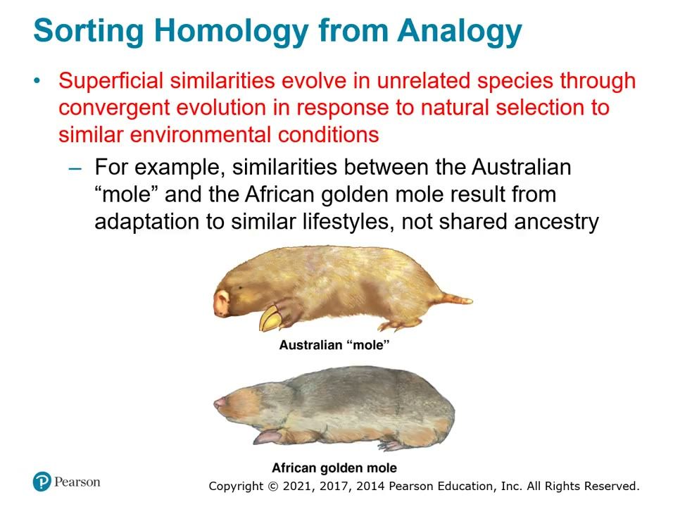
Slide 7. Analogy: Australian mole and African golden mole
The Australian "mole" and the African golden mole look alike because of convergent evolution to similar burrowing lifestyles, not shared ancestry. Their similarity is analogy, not homology, so it cannot be used to build a phylogeny.▶ Play from 4:04Lecture 26.2, slide 006
Be able to draw / explain
My notes
26.3

Shared characters are used to construct phylogenetic trees

Lecture video 26.3 (opens online)
Big idea
Cladistics groups organisms by common ancestry into clades, which are monophyletic (ancestor plus all descendants). Paraphyletic groups leave out some descendants, and polyphyletic groups leave out the most recent common ancestor. Trees are built from shared derived characters, identified by comparing an ingroup with an outgroup. Maximum parsimony picks the tree with the fewest evolutionary events, and maximum likelihood picks the tree most probable under rules of DNA change.

Must know

Terms

cladistics
An approach that classifies organisms by common ancestry.
clade
A group made of an ancestral species and all of its descendants.
monophyletic
A group containing an ancestor and all of its descendants.
paraphyletic
A group containing an ancestor and some, but not all, of its descendants.
polyphyletic
A group of distantly related species that does not include their most recent common ancestor.
shared ancestral character
A character that originated in an ancestor of the taxon being studied.
shared derived character
An evolutionary novelty unique to a particular clade.
outgroup
A species or group closely related to, but not part of, the group being studied.
ingroup
The group of species being studied.
maximum parsimony
The principle that the best tree needs the fewest evolutionary events (fewest base changes for DNA).
maximum likelihood
The principle that the best tree is the one most likely to produce the data, given probability rules for DNA change.

Examples

What she stressed in lecture
Memory hooks

Slides

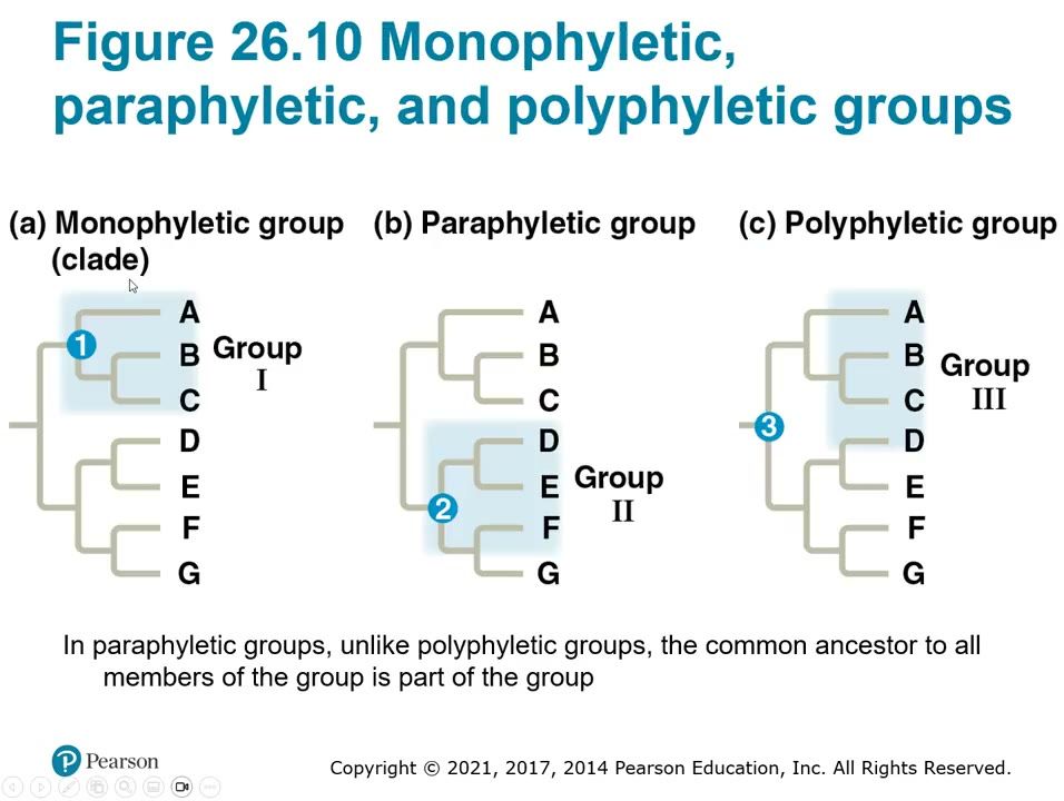
Slide 8. Monophyletic, paraphyletic and polyphyletic groups
A monophyletic group (clade) holds an ancestor and all its descendants; a paraphyletic group holds an ancestor and only some descendants; a polyphyletic group holds taxa with different ancestors. In a paraphyletic group, unlike a polyphyletic group, the common ancestor of all members is part of the group.
How to draw it: Draw three copies of a tree with tips A through G and shade Group I as a clade (ancestor 1 plus A, B, C), Group II as paraphyletic (ancestor 2 plus D, E, F, leaving out G), and Group III as polyphyletic (A, B, C, D without their shared ancestor 3); label each type.
▶ Play from 2:46Lecture 26.3, slide 006
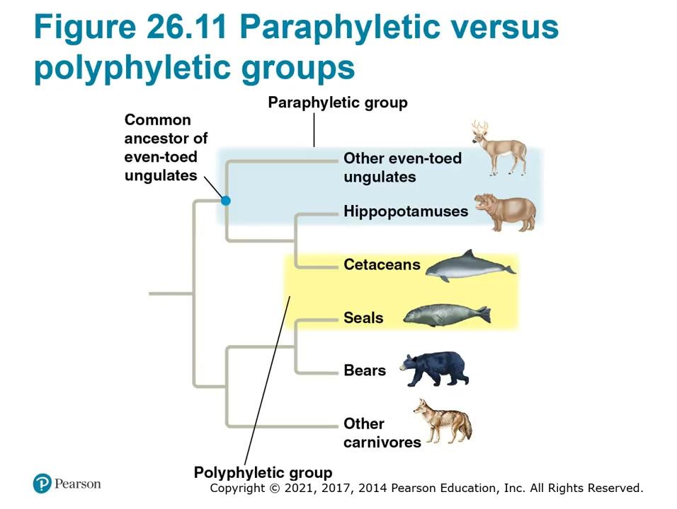
Slide 9. Even-toed ungulates and marine mammals
Grouping other even-toed ungulates with hippopotamuses but leaving out cetaceans makes a paraphyletic group. Grouping cetaceans with seals makes a polyphyletic group, because their similar body forms come from convergent evolution.▶ Play from 6:06Lecture 26.3, slide 007
Slide 10. Character table and tree from shared derived characters
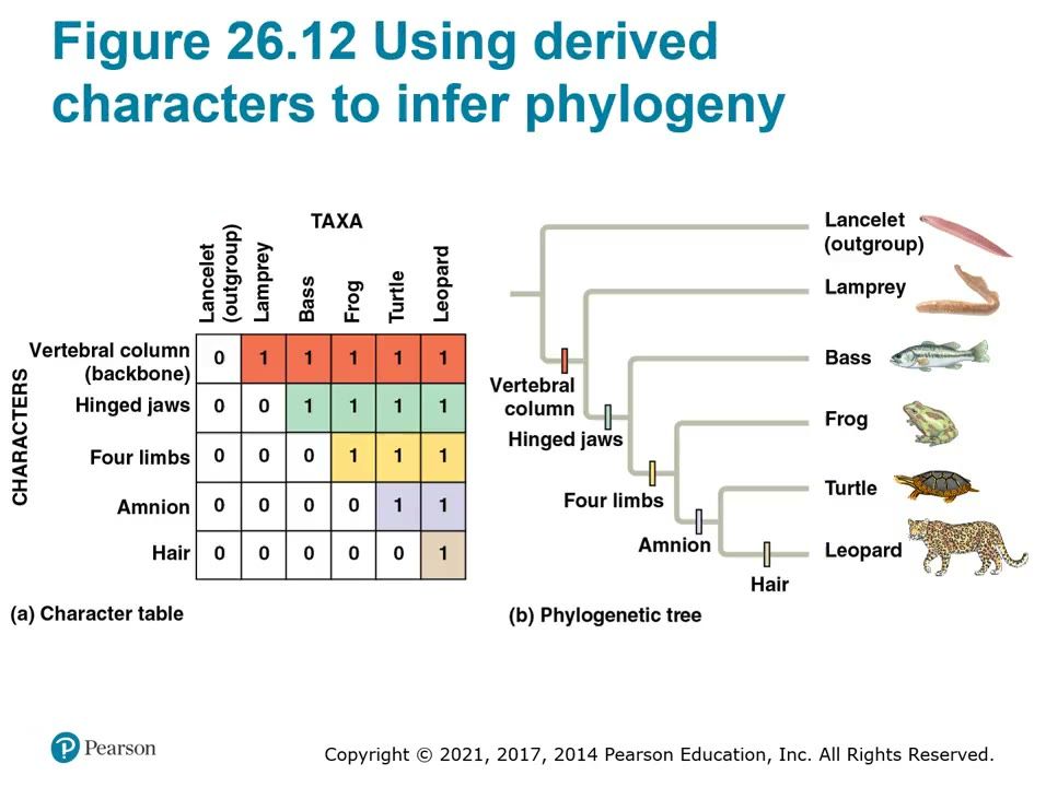
A 0/1 character table uses the lancelet as the outgroup to sort shared ancestral from shared derived characters. Each derived character (vertebral column, hinged jaws, four limbs, amnion, hair) appears once on the tree and marks the clade that has it.
How to draw it: Draw the character table (taxa lancelet, lamprey, bass, frog, turtle, leopard; characters vertebral column, hinged jaws, four limbs, amnion, hair) and the matching tree with a hash mark for each derived character on the branch where it first appears; label the lancelet as the outgroup.
▶ Play from 12:40Lecture 26.3, slide 013
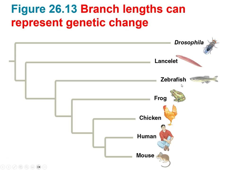
Slide 11. Branch lengths represent genetic change
In this tree, branch length shows the amount of genetic change in each lineage. The mouse branch is longer than the human branch, so the mouse lineage has changed more even though both have had the same time since their common ancestor.▶ Play from 16:56Lecture 26.3, slide 018
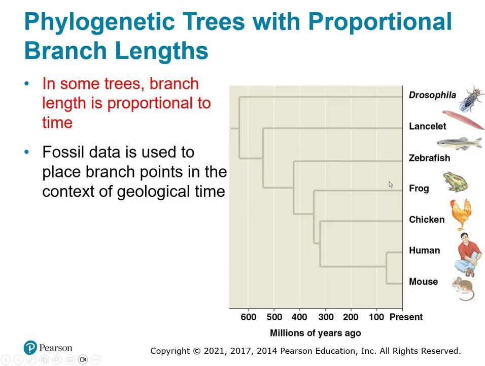
Slide 12. Branch lengths proportional to time
Here branch lengths are drawn to a time scale in millions of years ago, so all tips end at the present. Fossil data places the branch points in geological time.▶ Play from 16:56Lecture 26.3, slide 019
Be able to draw / explain
My notes
26.4

An organism's evolutionary history is documented in its genome

Lecture video 26.4 (opens online)
Big idea
Molecular comparisons reveal relatedness among groups that are morphologically dissimilar or poorly represented in the fossil record. Different genes evolve at different rates: rRNA genes change slowly (ancient relationships) and mitochondrial DNA changes fast (recent events). Gene duplication creates gene families, and homologous genes are either orthologous (from speciation) or paralogous (from duplication within a species).

Must know

Terms

gene duplication
Copying of a gene that increases the number of genes in a genome.
gene family
A group of related genes within a genome produced by repeated gene duplications.
orthologous genes
Homologous genes in different species whose homology results from speciation.
paralogous genes
Homologous genes within one species whose homology results from gene duplication.

Examples

What she stressed in lecture
Memory hooks

Slides

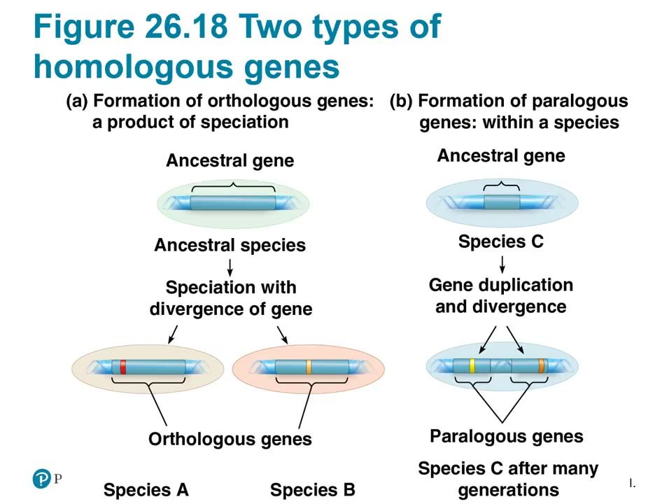
Slide 13. Orthologous vs paralogous genes
Orthologous genes form when speciation splits an ancestral gene between species A and species B. Paralogous genes form when a gene duplicates and the copies diverge within one species; orthologs are the ones used to infer phylogeny.
How to draw it: Draw (a) an ancestral gene in an ancestral species splitting by speciation into species A and species B, labeled orthologous genes; and (b) an ancestral gene in species C duplicating into two diverged copies in the same genome, labeled paralogous genes.
▶ Play from 3:25Lecture 26.4, slide 008
Be able to draw / explain
My notes
26.5

Molecular clocks help track evolutionary time

Lecture video 26.5 (opens online)
Big idea
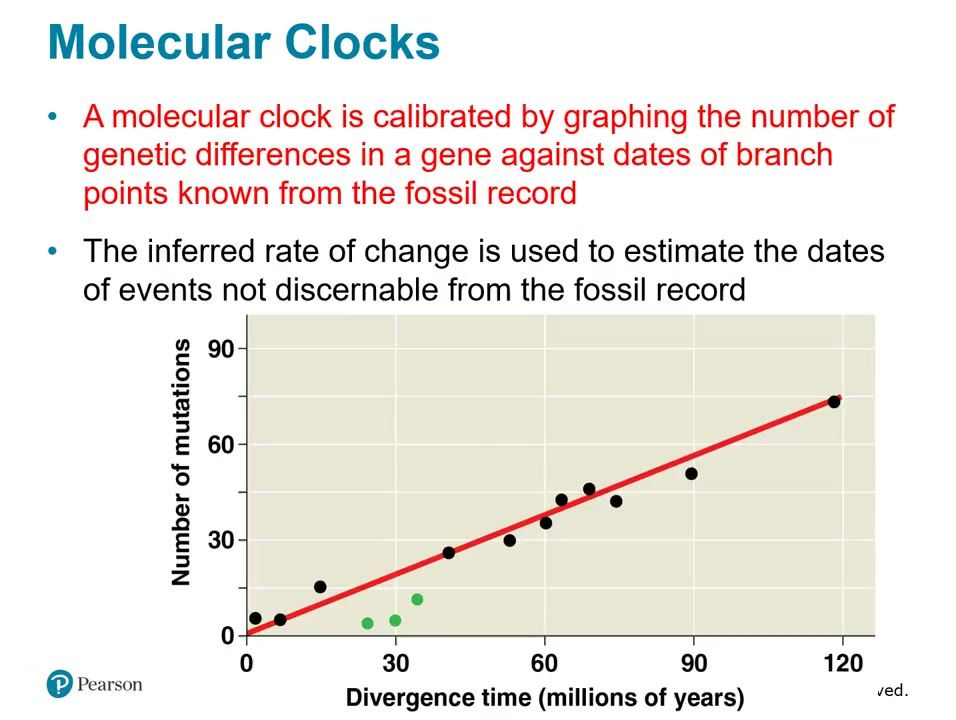
A molecular clock estimates the absolute time of evolutionary change, based on the observation that some genes evolve at a constant rate. It assumes the number of nucleotide substitutions is proportional to time since the split from a common ancestor (orthologs) or since gene duplication (paralogs). Clocks are calibrated against branch-point dates from the fossil record, and they have limits because some genes change in bursts and rates differ among genes and taxa.

Must know

Terms

molecular clock
A method that uses constant rates of molecular change to estimate the absolute time of evolutionary events.
neutral mutation
A mutation that is neither beneficial nor detrimental.
calibration
Setting the clock rate by graphing genetic differences against fossil-dated branch points.

Examples

What she stressed in lecture
Memory hooks

Slides

Slide 14. Calibrating a molecular clock
The graph plots number of mutations in a gene against divergence time known from fossils; the straight line gives the rate of change. That rate estimates dates of branch points not found in the fossil record.
How to draw it: Draw a graph with number of mutations on the y axis and divergence time (millions of years) on the x axis, points from fossil-dated branch points, and a best-fit straight line; label the line as the molecular clock rate.
▶ Play from 2:27Lecture 26.5, slide 004
Be able to draw / explain
My notes
26.6

Our understanding of the tree of life continues to change based on new data

Lecture video 26.6 (opens online)
Big idea
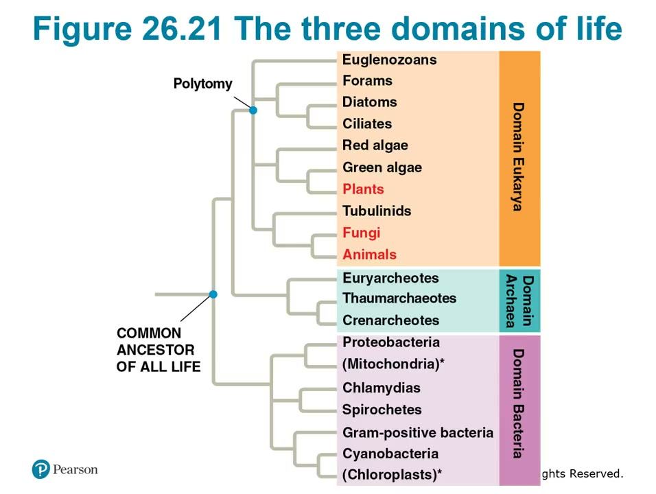
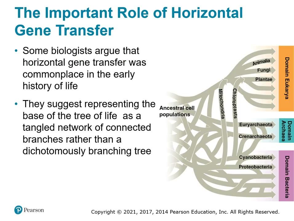
Classification moved from two kingdoms (plants and animals) to five kingdoms by the 1960s (Monera, Protista, Plantae, Fungi, Animalia) to the current three domains (Bacteria, Archaea, Eukarya). Prokaryotes form two domains because some differ from each other as much as from eukaryotes, and Archaea are more closely related to Eukarya than to Bacteria. Horizontal gene transfer moved genes between genomes and even between domains, so the early tree of life looks more like a tangled web than a clean branching tree.
Must know

Terms

domain
The broadest taxonomic category, above kingdom: Bacteria, Archaea, Eukarya.
Monera
Former kingdom that held all prokaryotes in the five-kingdom system.
horizontal gene transfer
Movement of genes from one genome to another, between species or even domains.
Examples

What she stressed in lecture
Memory hooks

Slides

Slide 15. The three domains of life
The rRNA-based tree splits life into Bacteria, Archaea and Eukarya, with Archaea and Eukarya more closely related to each other than either is to Bacteria. Mitochondria came from proteobacteria and chloroplasts from cyanobacteria; the polytomy marks unresolved branching.
How to draw it: Draw a tree from the common ancestor of all life with three branches labeled Domain Bacteria, Domain Archaea and Domain Eukarya, with Archaea and Eukarya as sister groups; mark plants, fungi and animals inside Eukarya.
▶ Play from 3:25Lecture 26.6, slide 006
Slide 16. Tangled base of the tree of life
Horizontal gene transfer was common early in the history of life, so some biologists draw the base of the tree as a tangled network rather than a dichotomously branching tree. The mitochondria and chloroplasts arrows show genes moving from bacteria into eukaryotes.▶ Play from 8:01Lecture 26.6, slide 011
Be able to draw / explain
My notes
27.1

Structural and functional adaptations contribute to prokaryotic success

Lecture video 27.1 (opens online)
Big idea
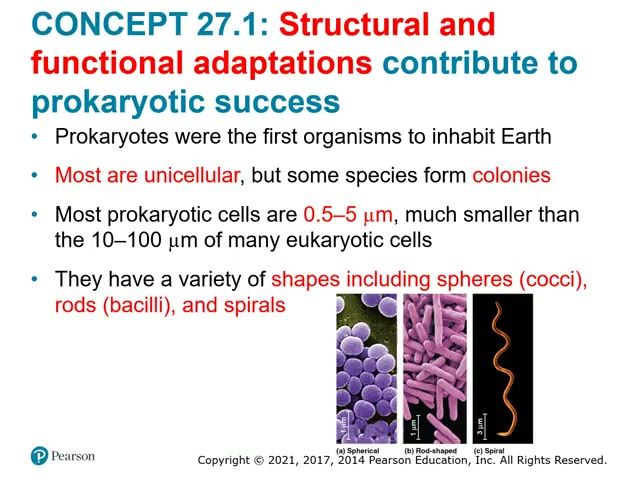
Prokaryotes (domains Bacteria and Archaea) were the first organisms on Earth and are the most abundant. They are small, mostly unicellular, and come as cocci, bacilli, and spirals. Surface structures (cell wall, capsule, fimbriae, pili, endospores) and flagella help them survive; they lack membrane-bound organelles and carry one circular chromosome in a nucleoid, plus plasmids. They reproduce by binary fission with short generation times.

Must know

Terms

prokaryote
Single-celled organism without a nucleus or membrane-bound organelles; domains Bacteria and Archaea.
cocci / bacilli / spirals
Sphere-shaped / rod-shaped / spiral-shaped prokaryotes.
peptidoglycan
Bacterial cell wall material made of sugars cross-linked by polypeptides.
Gram stain
A stain that classifies bacteria as Gram-positive or Gram-negative by cell wall composition.
Gram-positive
Bacteria with a thick peptidoglycan wall; stain purple.
Gram-negative
Bacteria with a thin peptidoglycan layer and an outer lipopolysaccharide membrane; stain pink/red; more antibiotic resistant.
capsule
A dense, sticky polysaccharide or protein layer around the cell wall.
endospore
A dormant, resistant cell formed inside a bacterium to survive harsh conditions.
fimbriae
Short hairlike projections for attaching to surfaces or other cells.
pili (sex pili)
Longer appendages that connect two cells for DNA transfer.
taxis
Movement toward or away from a stimulus.
nucleoid
Region of a prokaryotic cell that holds the circular chromosome, with no membrane around it.
plasmid
Small ring of DNA that replicates independently of the chromosome.
binary fission
Asexual division of a prokaryotic cell into two identical cells.

Examples

What she stressed in lecture
Memory hooks

Slides

Slide 17. Prokaryote shapes: cocci, bacilli, spirals
Prokaryotes come in three common shapes: spheres (cocci), rods (bacilli) and spirals. Most prokaryotic cells are 0.5 to 5 micrometers, much smaller than the 10 to 100 micrometers of many eukaryotic cells.▶ Play from 0:19Lecture 27.1, slide 002
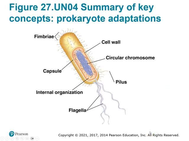
Slide 18. Prokaryote adaptations summary
A labeled bacterium shows the structures behind prokaryotic success: fimbriae, capsule, cell wall, circular chromosome, pilus, internal organization and flagella. Know the function of each part.
How to draw it: Draw a rod-shaped bacterium and label the capsule, cell wall, circular chromosome, internal organization, fimbriae, pilus and flagella.
▶ Play from 1:55Lecture 27.1, slide 003
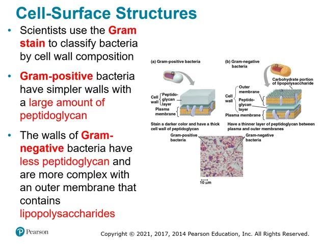
Slide 19. Gram-positive vs gram-negative cell walls
Gram-positive walls have a thick peptidoglycan layer and stain darker. Gram-negative walls have a thin peptidoglycan layer between the plasma membrane and an outer membrane with lipopolysaccharides, and they resist antibiotics more.
How to draw it: Draw both walls in cross section. Gram-positive: plasma membrane under a thick peptidoglycan layer. Gram-negative: plasma membrane, thin peptidoglycan layer, then outer membrane with lipopolysaccharide; label each layer.
▶ Play from 5:12Lecture 27.1, slide 006
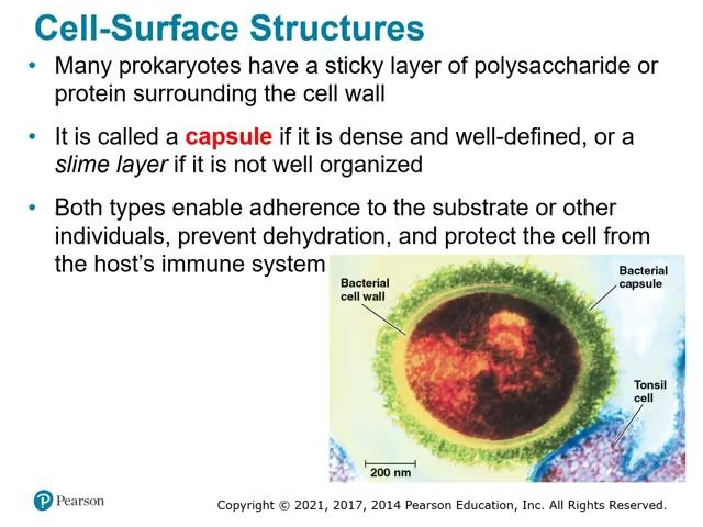
Slide 20. Bacterial capsule
A dense, well-defined sticky layer of polysaccharide or protein outside the cell wall is a capsule (a loose one is a slime layer). It lets the cell stick to surfaces such as this tonsil cell, prevents dehydration and protects against the host immune system.▶ Play from 8:25Lecture 27.1, slide 008
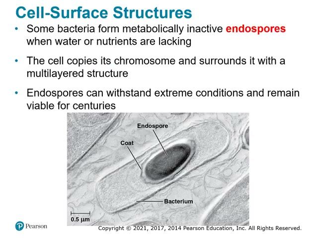
Slide 21. Endospore
When water or nutrients run out, some bacteria copy the chromosome and wrap it in a multilayered coat to form a metabolically inactive endospore. Endospores withstand extreme conditions and stay viable for centuries.▶ Play from 9:51Lecture 27.1, slide 009
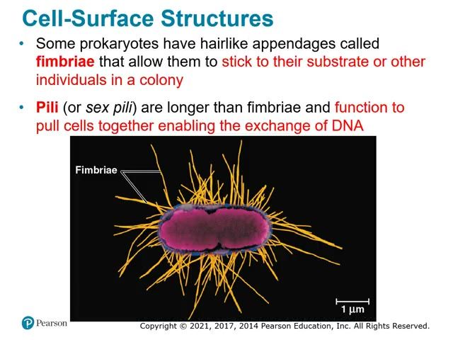
Slide 22. Fimbriae
Fimbriae are short hairlike appendages that let prokaryotes stick to a substrate or to each other in a colony. Pili (sex pili) are longer and pull cells together to exchange DNA.▶ Play from 11:33Lecture 27.1, slide 010
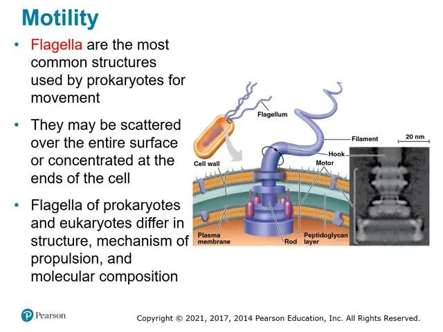
Slide 23. Prokaryotic flagellum
The bacterial flagellum has a filament, a hook and a motor anchored in the cell wall and plasma membrane by a rod. Prokaryotic and eukaryotic flagella differ in structure, mechanism of propulsion and molecular composition.
How to draw it: Draw the flagellum crossing the cell wall, peptidoglycan layer and plasma membrane; label the filament, hook, motor and rod.
▶ Play from 13:08Lecture 27.1, slide 012
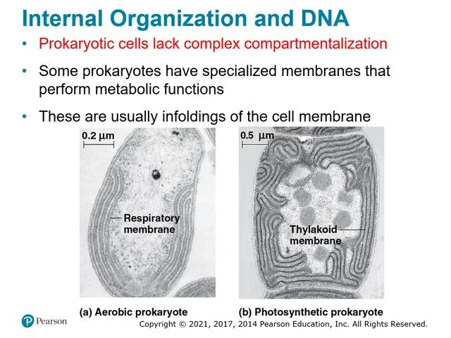
Slide 24. Specialized internal membranes
Prokaryotes lack complex compartments, but some have infoldings of the cell membrane that do metabolic work. An aerobic prokaryote has respiratory membranes and a photosynthetic prokaryote has thylakoid membranes.▶ Play from 14:09Lecture 27.1, slide 013
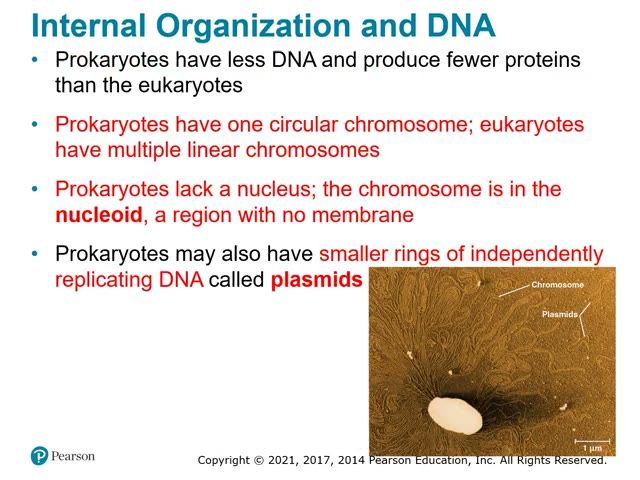
Slide 25. Chromosome and plasmids
A burst cell spills one long circular chromosome and several small rings of independently replicating DNA called plasmids. The chromosome sits in the nucleoid, a region with no membrane.▶ Play from 15:41Lecture 27.1, slide 014
Be able to draw / explain
My notes
27.2

Rapid reproduction, mutation, and genetic recombination promote genetic diversity in prokaryotes

Lecture video 27.2 (opens online)
Big idea
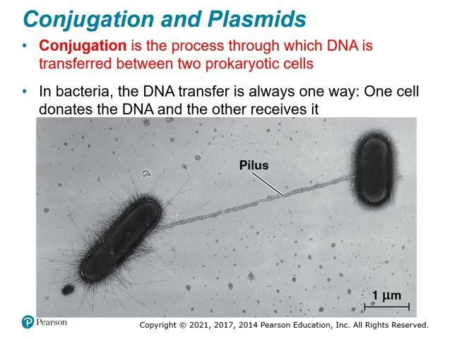
Three factors produce the high genetic diversity of prokaryotes: rapid reproduction, mutation, and genetic recombination. Binary fission makes identical cells, but mutations accumulate quickly because generations are short and populations are huge. Recombination combines DNA from two sources by transformation (DNA from the surroundings), transduction (phages carry genes), and conjugation (one-way DNA transfer between cells through a pilus).

Must know

Terms

genetic recombination
Combining DNA from two sources.
transformation
Uptake of foreign DNA from the surroundings.
transduction
Transfer of prokaryotic genes between cells by bacteriophages.
bacteriophage (phage)
A virus that infects bacteria.
conjugation
One-way transfer of DNA between two prokaryotic cells that are temporarily joined.

Examples

What she stressed in lecture
Memory hooks

Slides

Slide 26. Conjugation through a pilus
In conjugation, a pilus pulls two cells together and DNA passes one way from the donor cell to the recipient. R plasmids move this way and spread antibiotic resistance genes.▶ Play from 4:33Lecture 27.2, slide 007
Be able to draw / explain
My notes
27.3

Diverse nutritional and metabolic adaptations have evolved in prokaryotes

Lecture video 27.3 (opens online)
Big idea
Prokaryotes are classified by energy source (phototrophs use light, chemotrophs use chemicals) and carbon source (autotrophs use CO2, heterotrophs need organic nutrients), giving four nutritional modes. They differ in oxygen use (obligate aerobes, obligate anaerobes, facultative anaerobes). Some fix atmospheric nitrogen into ammonia, as in the heterocysts of Anabaena, and many cooperate in biofilms.

Must know

Terms

phototroph
Organism that obtains energy from light.
chemotroph
Organism that obtains energy from chemicals.
autotroph
Organism that uses CO2 or related compounds as its carbon source.
heterotroph
Organism that needs an organic nutrient as its carbon source.
photoautotroph
Uses light for energy and CO2 for carbon.
chemoautotroph
Uses inorganic chemicals for energy and CO2 for carbon.
photoheterotroph
Uses light for energy and organic compounds for carbon.
chemoheterotroph
Uses organic compounds for both energy and carbon.
obligate aerobe
Requires oxygen for cellular respiration.
obligate anaerobe
Cannot use oxygen and is poisoned by it.
facultative anaerobe
Uses oxygen when present, switches to fermentation or anaerobic respiration when it is absent.
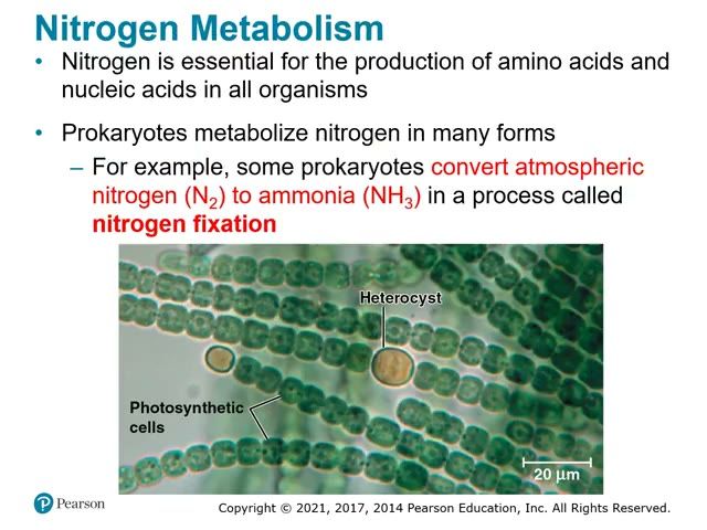
nitrogen fixation
Conversion of atmospheric nitrogen (N2) to ammonia (NH3).
heterocyst
Specialized cyanobacterial cell that fixes nitrogen.
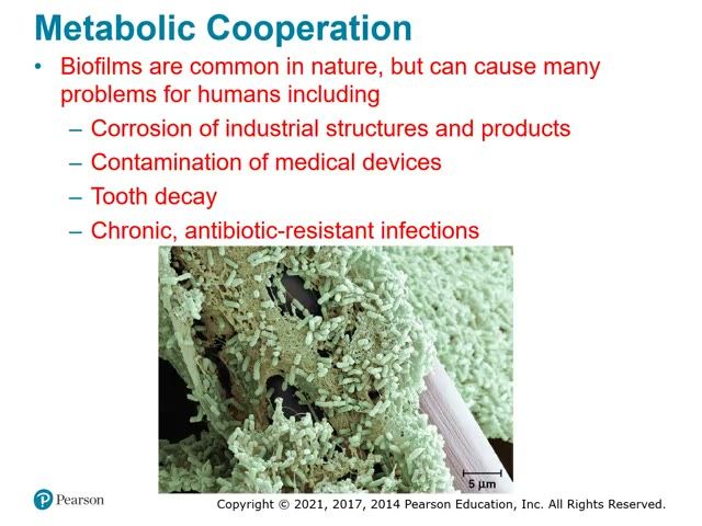
biofilm
A surface-coating colony formed by cooperating prokaryotes.

Examples

What she stressed in lecture
Memory hooks

Slides

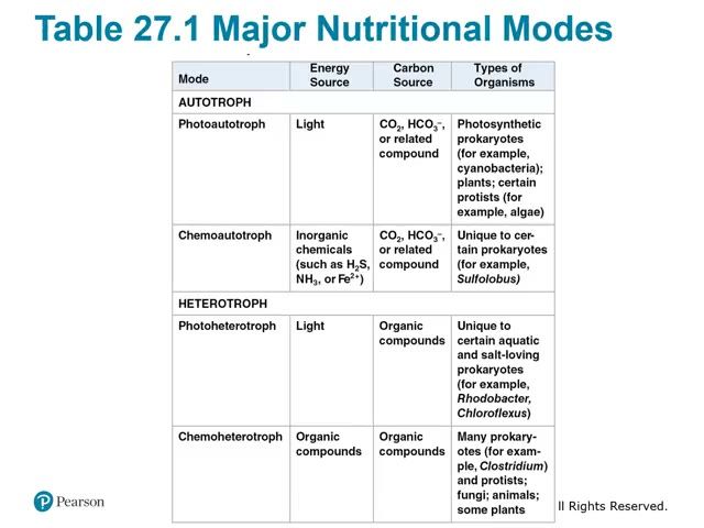
Slide 27. Table 27.1 Major nutritional modes
Energy source (light or chemicals) plus carbon source (CO2 or organic compounds) gives four modes: photoautotroph, chemoautotroph, photoheterotroph and chemoheterotroph. Chemoautotrophs (for example Sulfolobus) and photoheterotrophs are found only in certain prokaryotes; animals are chemoheterotrophs.▶ Play from 2:03Lecture 27.3, slide 004
Slide 28. Nitrogen fixation in Anabaena heterocysts
In the cyanobacterium Anabaena, most cells in the filament photosynthesize while heterocysts carry out nitrogen fixation, converting N2 to NH3. The heterocyst shares fixed nitrogen with the other cells, an example of metabolic cooperation.▶ Play from 4:40Lecture 27.3, slide 006
Slide 29. Biofilm
A biofilm is a surface-coating colony of cooperating prokaryotes, with channels that bring in nutrients and remove wastes. Biofilms cause corrosion, contaminate medical devices, cause tooth decay and cause chronic antibiotic-resistant infections.▶ Play from 7:36Lecture 27.3, slide 008
Be able to draw / explain
My notes
27.4

Prokaryotes have radiated into a diverse set of lineages

Lecture video 27.4 (opens online)
Big idea
Prokaryotes date to about 3.5 billion years ago and inhabit every environment that supports life. Genetic analysis in the 1970s split them into Bacteria and Archaea. Bacteria include Proteobacteria, Chlamydias, Spirochetes, Cyanobacteria (all Gram-negative) and Gram-positive bacteria. Archaea share traits with both bacteria and eukaryotes and include extremophiles (extreme halophiles, extreme thermophiles) and methanogens, organized into Euryarchaeota, the TACK supergroup, and Lokiarchaeotes.

Must know

Terms

metagenomics
Studying all the prokaryotic genomes in an environmental sample to identify which species are present.
Proteobacteria
Large Gram-negative bacterial clade with photoautotrophs, chemoautotrophs, and heterotrophs.
Chlamydias
Gram-negative animal-cell parasites whose walls lack peptidoglycan.
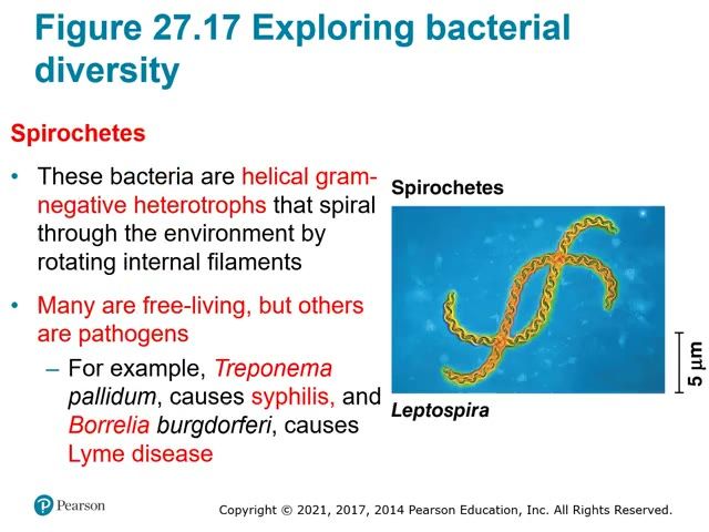
Spirochetes
Helical Gram-negative heterotrophs that move by rotating internal filaments.
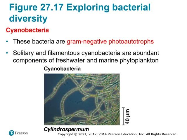
Cyanobacteria
Gram-negative photoautotrophic bacteria.
Actinomycetes
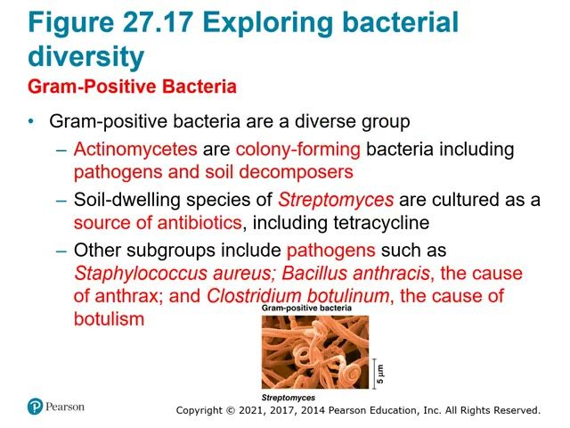
Colony-forming Gram-positive bacteria that include pathogens and soil decomposers.
extremophiles
Archaea that live in extreme environments uninhabitable for most organisms.
extreme halophiles
Archaea that tolerate or require very salty environments.
extreme thermophiles
Archaea adapted to very high temperatures.
methanogens
Obligately anaerobic archaea that produce methane.
TACK supergroup
Group of closely related archaeal clades: Thaumarchaeota, Aigarchaeota, Crenarchaeota, Korarchaeota.

Examples

What she stressed in lecture
Memory hooks

Slides

Slide 30. Simplified phylogeny of prokaryotes
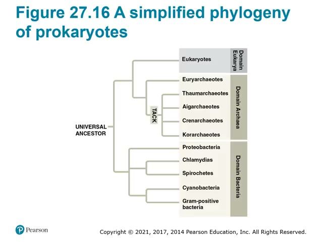
From the universal ancestor, Domain Bacteria splits from the line leading to Archaea and Eukarya. Archaea includes the Euryarchaeotes and the TACK supergroup (Thaumarchaeotes, Aigarchaeotes, Crenarchaeotes, Korarchaeotes).▶ Play from 3:08Lecture 27.4, slide 004
Slide 31. Known vs undiscovered bacterial species
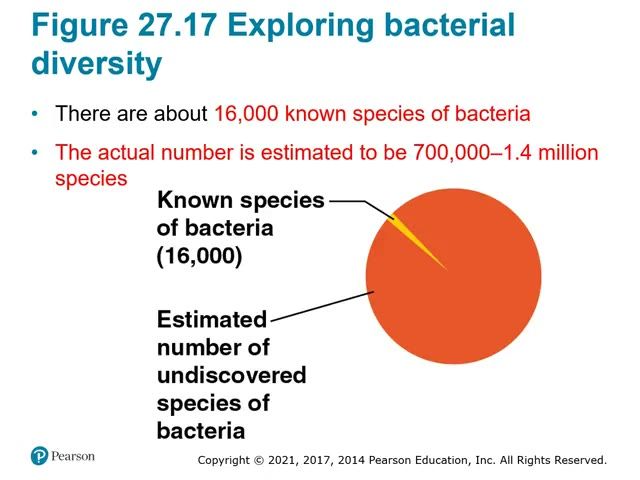
About 16,000 bacterial species are named, a thin slice of the estimated 700,000 to 1.4 million species. Metagenomics now reveals much of this unseen diversity.▶ Play from 6:03Lecture 27.4, slide 008
Slide 32. Proteobacteria: Thiomargarita namibiensis
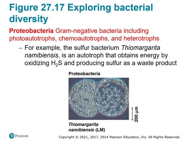
Proteobacteria are gram-negative and include photoautotrophs, chemoautotrophs and heterotrophs. Thiomargarita namibiensis is a sulfur bacterium that gets energy by oxidizing H2S and releases sulfur as waste.▶ Play from 6:39Lecture 27.4, slide 011
Slide 33. Chlamydias: Chlamydia inside an animal cell
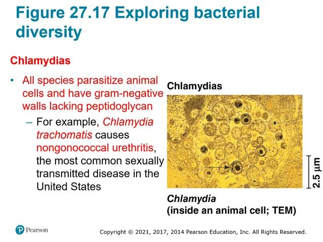
All chlamydias parasitize animal cells and have gram-negative walls lacking peptidoglycan. Chlamydia trachomatis causes nongonococcal urethritis, the most common sexually transmitted disease in the United States.▶ Play from 8:23Lecture 27.4, slide 013
Slide 34. Spirochetes: Leptospira
Spirochetes are helical gram-negative heterotrophs that spiral by rotating internal filaments. Pathogens include Treponema pallidum (syphilis) and Borrelia burgdorferi (Lyme disease).▶ Play from 9:03Lecture 27.4, slide 014
Slide 35. Cyanobacteria: Cylindrospermum
Cyanobacteria are gram-negative photoautotrophs that make up much of freshwater and marine phytoplankton, as solitary cells or filaments. Chloroplasts descend from cyanobacteria.▶ Play from 10:19Lecture 27.4, slide 015
Slide 36. Gram-positive bacteria: Streptomyces
Gram-positive bacteria include colony-forming actinomycetes; soil-dwelling Streptomyces is the source of antibiotics such as tetracycline. Gram-positive pathogens include Staphylococcus aureus, Bacillus anthracis (anthrax) and Clostridium botulinum (botulism).▶ Play from 11:01Lecture 27.4, slide 016
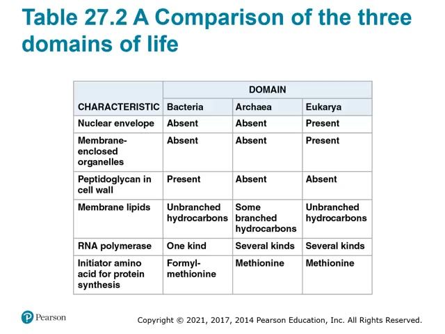
Slide 37. Table 27.2 Comparison of the three domains
Bacteria and Archaea both lack a nuclear envelope and membrane-enclosed organelles; only Bacteria have peptidoglycan. Archaea match Eukarya in having several kinds of RNA polymerase and methionine as the initiator amino acid, while Bacteria use formyl-methionine.▶ Play from 13:44Lecture 27.4, slide 019
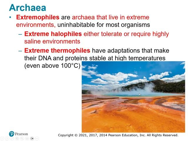
Slide 38. Extreme thermophiles: hot spring
Extremophile archaea live where most organisms cannot: extreme halophiles need very salty water, and extreme thermophiles like those in this hot spring have DNA and proteins stable above 100 degrees C. Most extreme thermophiles belong to the Crenarchaeota in the TACK supergroup.▶ Play from 16:33Lecture 27.4, slide 024
Be able to draw / explain
My notes
27.5

Prokaryotes play crucial roles in the biosphere

Lecture video 27.5 (opens online)
Big idea
Without prokaryotes, the prospects for other life on Earth would be dim. They recycle chemical elements between living and nonliving parts of the environment, act as decomposers, fix CO2 and nitrogen into forms other organisms use, and can also immobilize nutrients. They form symbioses (mutualism, commensalism, parasitism) with larger hosts, and some ecosystems such as hydrothermal vents depend on them.

Must know

Terms

decomposer
Organism that breaks down dead organisms and wastes and returns elements to the environment.
symbiosis
An ecological relationship in which two species live in close contact.
host
The larger organism in a symbiosis.
symbiont
The smaller organism in a symbiosis.
mutualism
Symbiosis in which both organisms benefit.
commensalism
Symbiosis in which one organism benefits and the other is neither helped nor harmed.
parasitism
Symbiosis in which the parasite harms, but usually does not kill, the host.
pathogen
A parasite that causes disease.

Examples

What she stressed in lecture
Memory hooks
My notes
27.6

Prokaryotes have both beneficial and harmful impacts on humans

Lecture video 27.6 (opens online)
Big idea
The best-known prokaryotes are pathogens, but they are a small fraction of all prokaryotes. Mutualistic gut bacteria make nutrients, and prokaryotes are used in food production, DNA technology, plastics, biofuels, and bioremediation. All known pathogenic prokaryotes are bacteria; they cause disease through exotoxins or endotoxins, and antibiotic resistance has evolved rapidly since antibiotics were introduced in the 1940s.

Must know

Terms

exotoxin
A protein secreted by bacteria that causes disease even after the bacteria are gone.
endotoxin
Lipopolysaccharide from the outer membrane of Gram-negative bacteria, released when the bacteria die.
antibiotic resistance
Evolved ability of bacteria to survive an antibiotic.
CRISPR-Cas9
A prokaryotic defense system against viruses, now used as a gene-editing tool.
bioremediation
Use of organisms to remove pollutants from soil, air, or water.

Examples

What she stressed in lecture
Memory hooks

Slides

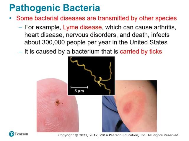
Slide 39. Lyme disease: tick, spirochete and rash
Lyme disease is caused by the spirochete Borrelia burgdorferi, carried to humans by ticks, and often shows a red rash at the bite. It can cause arthritis, heart disease, nervous disorders and death.▶ Play from 2:42Lecture 27.6, slide 006
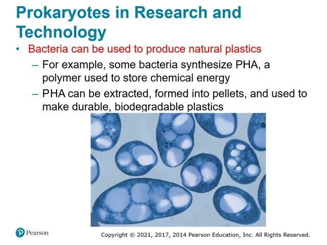
Slide 40. Bacteria making PHA plastic
Some bacteria store chemical energy as the polymer PHA, seen as the pale granules inside these cells. PHA is extracted, formed into pellets and made into durable, biodegradable plastics.▶ Play from 11:05Lecture 27.6, slide 015
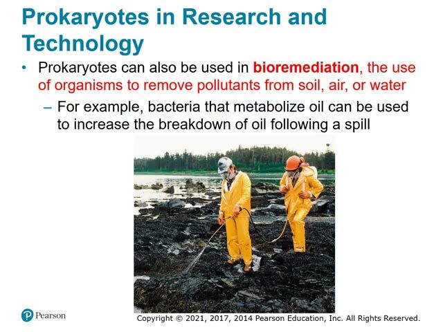
Slide 41. Bioremediation of an oil spill
Bioremediation uses organisms to remove pollutants from soil, air or water. After an oil spill, oil-metabolizing bacteria speed the breakdown of the oil.▶ Play from 11:55Lecture 27.6, slide 017
Be able to draw / explain
My notes
PV

Phylogenetics videos: taxonomy, homology vs analogy, and the anole lizards

Big idea
Crash Course 'Taxonomy' covers Linnaeus and the hierarchy, using the cat (Felis catus) as a walk-through. A short video shows that homologous structures come from common ancestry while analogous structures come from convergent evolution. The anole lizard video (Jonathan Losos with Sean Carroll) shows adaptation, rapid evolution, reproductive isolation, and DNA-based tree building that revealed the same body types evolved independently on each Caribbean island.

Must know

Terms

taxonomy
The science of classifying living things.
dewlap
A flap of throat skin that male anoles display to attract females.
reproductive isolation
Something that prevents members of one population from breeding with a closely related population.

Examples

What she stressed in lecture
Memory hooks
My notes
28.1

Most eukaryotes are single-celled organisms

Lecture video 28.1 (opens online)
Big idea
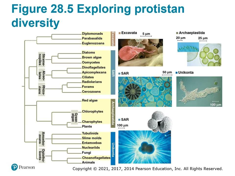
"Protist" is an informal term for all eukaryotes that are not plants, animals or fungi. Kingdom Protista no longer exists because some protists are more closely related to plants, fungi or animals than to other protists. Protists are the most structurally, functionally and nutritionally diverse eukaryotes. All eukaryotes, protists included, are sorted into four supergroups: Excavata, SAR, Archaeplastida and Unikonta (Figure 28.5).

Must know

Terms

protist
Informal name for any eukaryote that is not a plant, animal or fungus.
mixotroph
An organism that combines photosynthesis with heterotrophic nutrition.
supergroup
One of the four largest groupings of eukaryotes: Excavata, SAR, Archaeplastida, Unikonta.
major group
A large clade inside a supergroup (gray in Fig. 28.5), e.g. stramenopiles, green algae, amoebozoans.
pseudopodium (pl. pseudopodia)
Extension of the cell surface used to move and to engulf food.
phagocytosis
Engulfing food particles by surrounding them with the cell membrane.
ectoplasm / endoplasm
Clear, actin-rich outer cytoplasm at the amoeba's leading edge / the inner, granular cytoplasm that flows forward.

Examples

What she stressed in lecture
Memory hooks

Slides

Slide 42. Figure 28.5 Exploring protistan diversity (full tree)
The four supergroups from the common ancestor of eukaryotes: Excavata, SAR, Archaeplastida, Unikonta, with every clade and major group. Needed for lecture AND lab.
How to draw it: Draw a root with four branches; list the clades under each supergroup from memory.
▶ Play from 4:22Lecture 28.1, slide 007
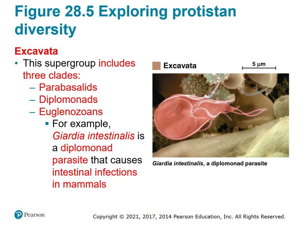
Slide 43. Excavata: Giardia intestinalis
Excavata has three clades (parabasalids, diplomonads, euglenozoans). Giardia is a diplomonad parasite causing intestinal infection in mammals.▶ Play from 7:31Lecture 28.1, slide 008
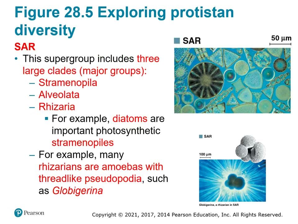
Slide 44. SAR: diatoms and the foram Globigerina
SAR has three major groups: stramenopiles, alveolates, rhizarians. Diatoms are important photosynthetic stramenopiles; Globigerina is a rhizarian with thread-like pseudopodia.▶ Play from 8:35Lecture 28.1, slide 009
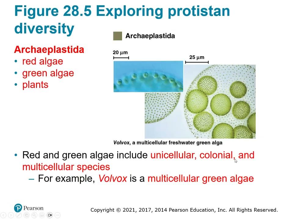
Slide 45. Archaeplastida: Volvox
Red algae, green algae, plants. Red and green algae include unicellular, colonial and multicellular species; Volvox is a multicellular freshwater green alga.▶ Play from 9:41Lecture 28.1, slide 010
Slide 46. Unikonta: Amoeba proteus
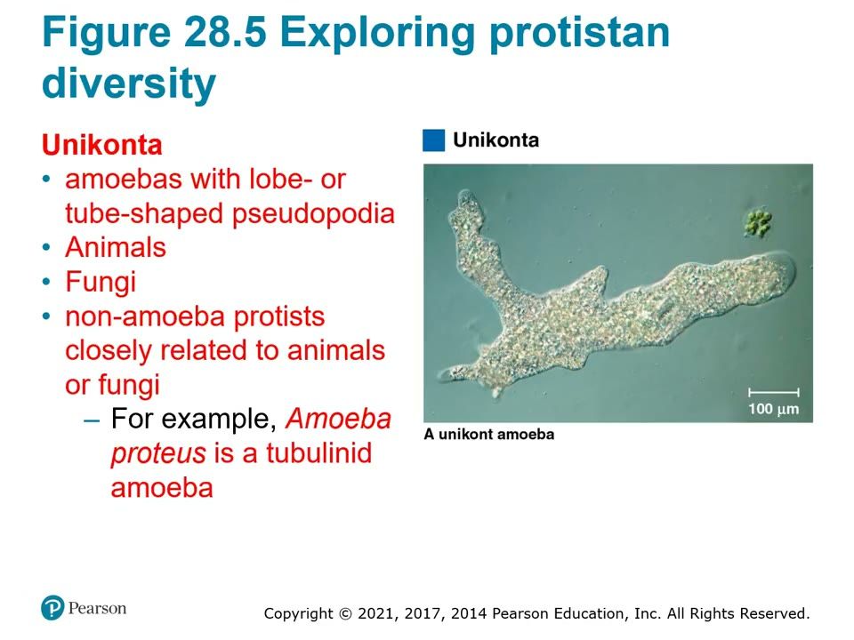
Amoebas with lobe- or tube-shaped pseudopodia, animals, fungi and protists close to them. Amoeba proteus is a tubulinid.
How to draw it: Amoeba outline with blunt lobe-shaped pseudopodia; label pseudopodium, ectoplasm, endoplasm.
▶ Play from 10:33Lecture 28.1, slide 013
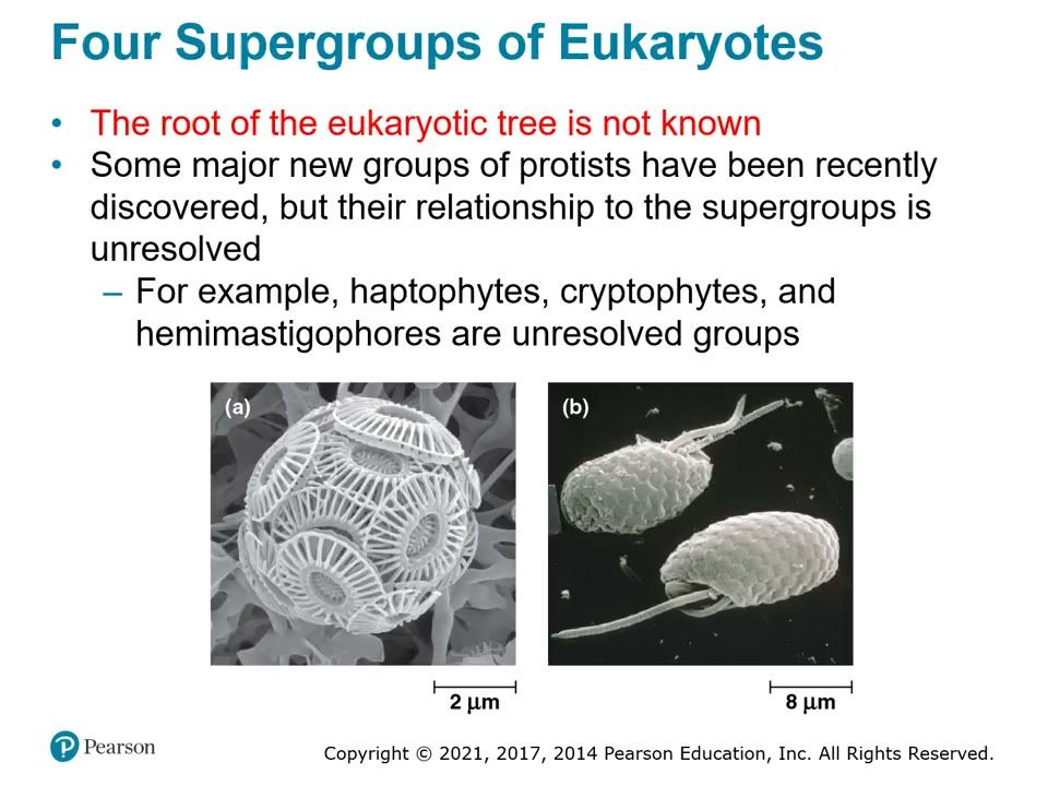
Slide 47. Unresolved groups
The root of the eukaryotic tree is not known; haptophytes, cryptophytes and hemimastigophores are not yet placed in a supergroup.▶ Play from 12:28Lecture 28.1, slide 020
Be able to draw / explain
My notes
28.2

Excavates include protists with modified mitochondria and protists with unique flagella

Lecture video 28.2 (opens online)
Big idea
Excavata is defined by its cytoskeleton; some members have an excavated feeding groove on one side of the body. It has three monophyletic groups (clades): diplomonads, parabasalids and euglenozoans. Diplomonads and parabasalids lack plastids, have reduced mitochondria and mostly live in anaerobic environments. Euglenozoans have a spiral or crystalline rod inside each flagellum and split into kinetoplastids and euglenids.

Must know

Terms

Excavata
Supergroup defined by its cytoskeleton; some have a feeding groove.
mitosome
Reduced mitochondrion of diplomonads; lacks an electron transport chain.
hydrogenosome
Reduced mitochondrion of parabasalids; makes some ATP anaerobically, releasing H2.
undulating membrane
Wavy membrane along the side of Trichomonas (and Trypanosoma) used in movement.
kinetoplast
Organized mass of DNA in the single large mitochondrion of a kinetoplastid.
pellicle
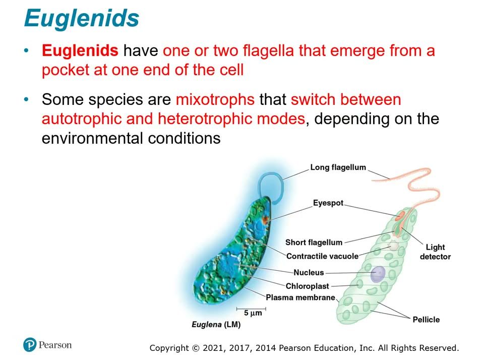
Flexible protein layer under the membrane of Euglena that lets it change shape.
eyespot
Red light-detecting spot near the flagellum base of Euglena.

Examples

What she stressed in lecture
Memory hooks

Slides

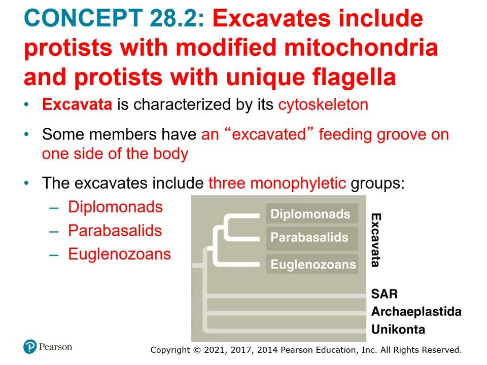
Slide 48. Excavata: three monophyletic groups
Diplomonads, parabasalids and euglenozoans. Excavata is defined by its cytoskeleton; some have an excavated feeding groove.▶ Play from 0:09Lecture 28.2, slide 002
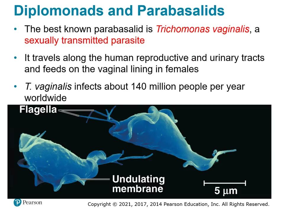
Slide 49. Trichomonas vaginalis (parabasalid)
Sexually transmitted parasite; flagella and the undulating membrane; about 140 million infections a year.
How to draw it: Teardrop cell, flagella at the front, a wavy undulating membrane down one side.
▶ Play from 3:30Lecture 28.2, slide 006
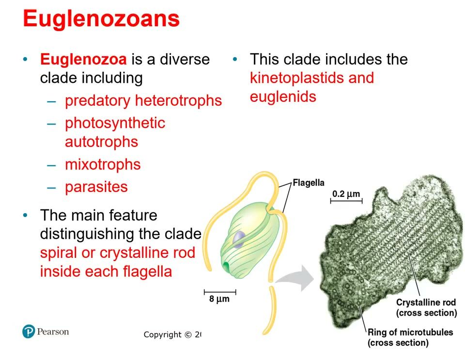
Slide 50. Euglenozoans: crystalline rod in the flagellum
Diverse clade (predatory heterotrophs, autotrophs, mixotrophs, parasites) with kinetoplastids and euglenids; defined by a spiral or crystalline rod inside each flagellum (ring of microtubules around it).▶ Play from 4:33Lecture 28.2, slide 007
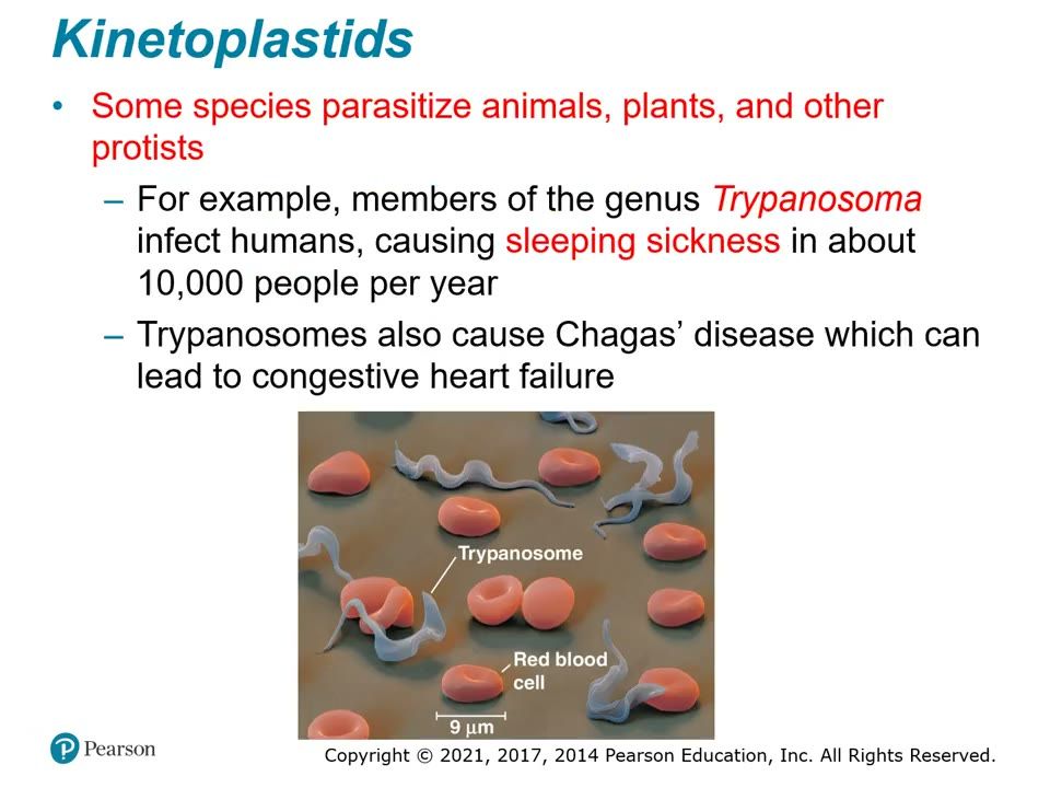
Slide 51. Trypanosoma among red blood cells
Kinetoplastid parasite: sleeping sickness (about 10,000 people a year) and Chagas disease (can cause congestive heart failure).▶ Play from 6:30Lecture 28.2, slide 009
Slide 52. Euglena, labeled
Euglenid: long and short flagella from a pocket at one end, eyespot and light detector, contractile vacuole, nucleus, chloroplasts, pellicle. Some are mixotrophs.
How to draw it: Spindle-shaped cell; long flagellum from the front pocket; red eyespot; chloroplast ovals; label pellicle.
▶ Play from 7:37Lecture 28.2, slide 010
Be able to draw / explain
My notes
28.3

The "SAR" clade is a highly diverse group of protists defined by DNA similarities

Lecture video 28.3 (opens online)
Big idea
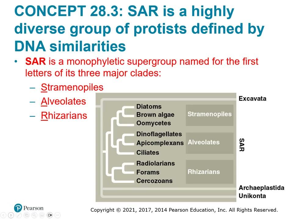
SAR is grouped by DNA similarity and has three major groups. Stramenopiles (hairy + smooth flagellum) include diatoms, brown algae and oomycetes. Alveolates (alveoli, sacs under the plasma membrane) include dinoflagellates, apicomplexans and ciliates. Rhizarians are mostly amoebas with thread-like pseudopodia: radiolarians, forams and cercozoans. The Plasmodium (malaria) life cycle is the one life cycle to know.

Must know

Terms

SAR
Supergroup named for its major groups: Stramenopiles, Alveolates, Rhizarians.
phytoplankton
Photosynthetic plankton (e.g. diatoms, dinoflagellates).
holdfast / stipe / blade
Root-like anchor / stem-like stalk / leaf-like photosynthetic surface of a brown alga.
alginate
Gel-forming substance in brown algal cell walls; food thickener.
alveoli
Membrane-enclosed sacs just under the plasma membrane of alveolates.
sporozoite
Infectious cell of an apicomplexan.
merozoite
Plasmodium stage released from liver cells that infects red blood cells.
gametocyte
Plasmodium stage in red blood cells that becomes gametes inside the mosquito.
micronucleus / macronucleus
Small nuclei exchanged in conjugation / large nucleus that runs the cell (ciliates).
conjugation
Exchange of micronuclei between two ciliates; genetic variation without reproduction.
test
Porous shell of a foram (calcium carbonate).

Examples

What she stressed in lecture
Memory hooks

Slides

Slide 53. SAR tree
Stramenopiles (diatoms, brown algae, oomycetes), alveolates (dinoflagellates, apicomplexans, ciliates), rhizarians (radiolarians, forams, cercozoans).▶ Play from 0:08Lecture 28.3, slide 002
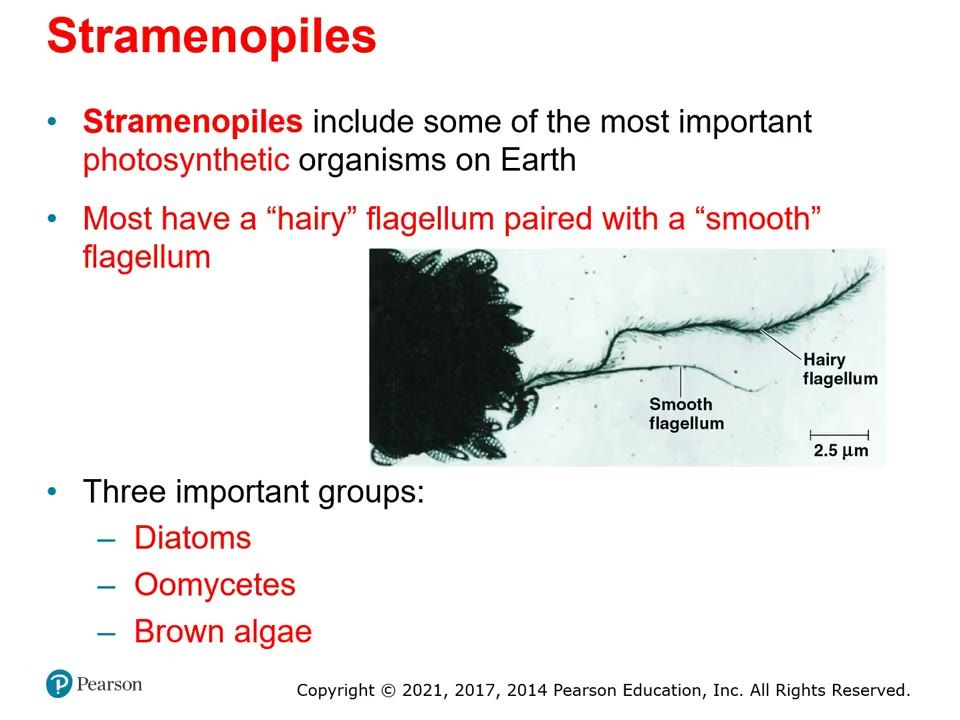
Slide 54. Stramenopile flagella
Most stramenopiles have a hairy flagellum paired with a smooth flagellum.▶ Play from 0:50Lecture 28.3, slide 003
Slide 55. Diatom
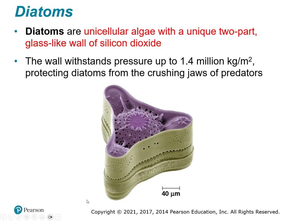
Unicellular alga with a two-part glass-like wall of silicon dioxide that withstands up to 1.4 million kg/m2.▶ Play from 1:35Lecture 28.3, slide 004
Slide 56. Brown alga body plan
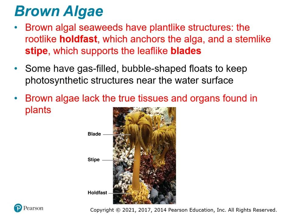
Root-like holdfast anchors; stem-like stipe supports leaf-like blades; some have gas-filled floats. No true tissues or organs.
How to draw it: Kelp: holdfast at the bottom, stipe, blades, a float; label all four.
▶ Play from 4:30Lecture 28.3, slide 008
Slide 57. Oomycetes (water molds)
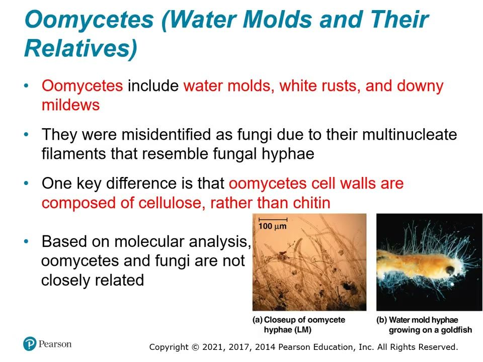
Water molds, white rusts, downy mildews. Filaments look like fungal hyphae, but walls are cellulose; not close relatives of fungi. Water mold growing on a goldfish.▶ Play from 6:37Lecture 28.3, slide 010
Slide 58. Alveoli
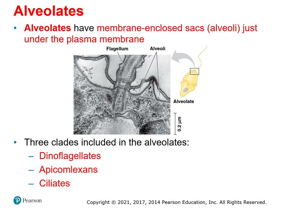
Alveolates have membrane-enclosed sacs (alveoli) just under the plasma membrane. Three clades: dinoflagellates, apicomplexans, ciliates.▶ Play from 8:58Lecture 28.3, slide 012
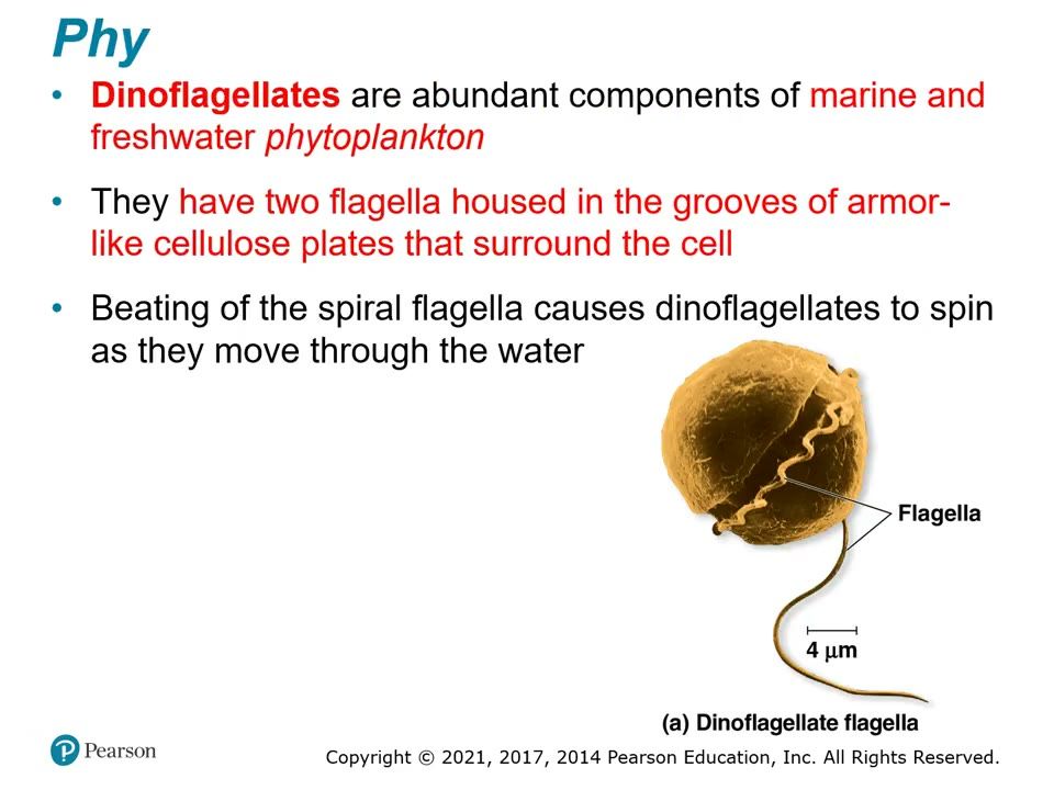
Slide 59. Dinoflagellate
Two flagella in grooves of armor-like cellulose plates; the spiral flagellum makes the cell spin.
How to draw it: Round cell with a groove around the middle and a second groove down; one flagellum in each.
▶ Play from 9:55Lecture 28.3, slide 013
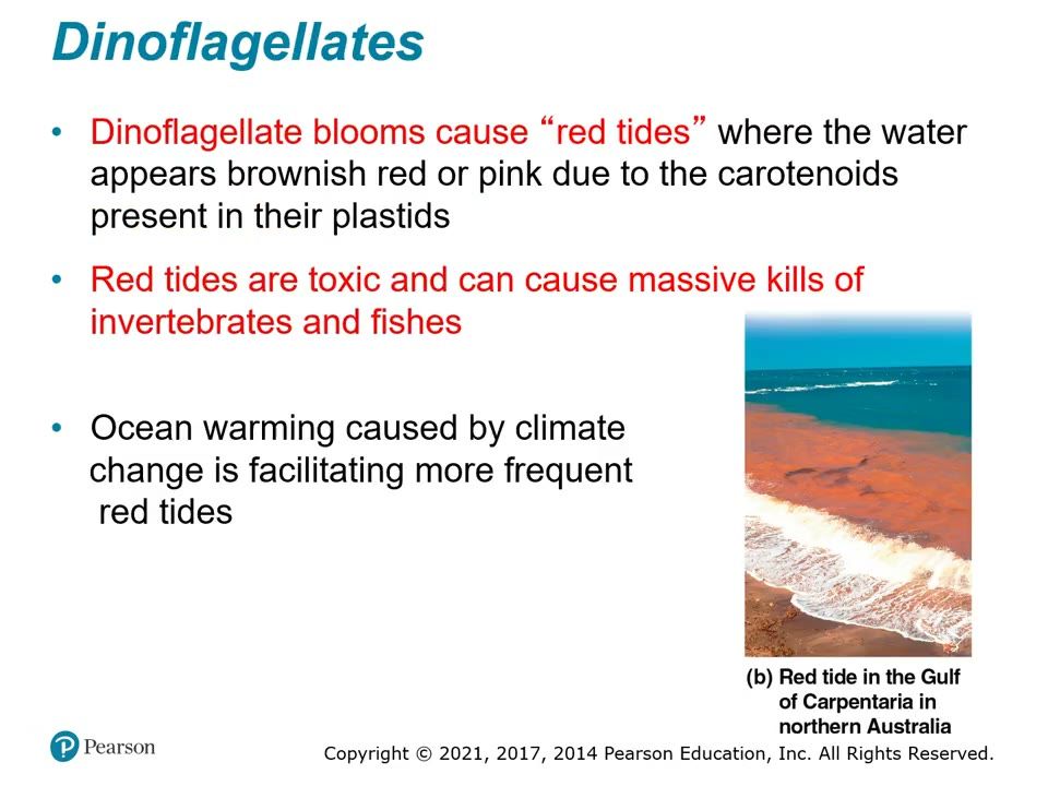
Slide 60. Red tide
Dinoflagellate bloom colors the water brownish red or pink (carotenoids); toxic; kills invertebrates and fish; becoming more frequent with ocean warming.▶ Play from 11:09Lecture 28.3, slide 016
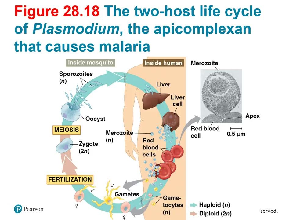
Slide 61. Figure 28.18 Plasmodium two-host life cycle
KNOW THIS (extra credit). Inside human: sporozoites (n) → liver cells → merozoites (n) → red blood cells → gametocytes (n). Inside mosquito: gametes → fertilization → zygote (2n) → meiosis → oocyst → sporozoites.
How to draw it: Two halves: mosquito (sexual) and human (asexual); label every stage with n or 2n; box MEIOSIS and FERTILIZATION.
▶ Play from 14:49Lecture 28.3, slide 019
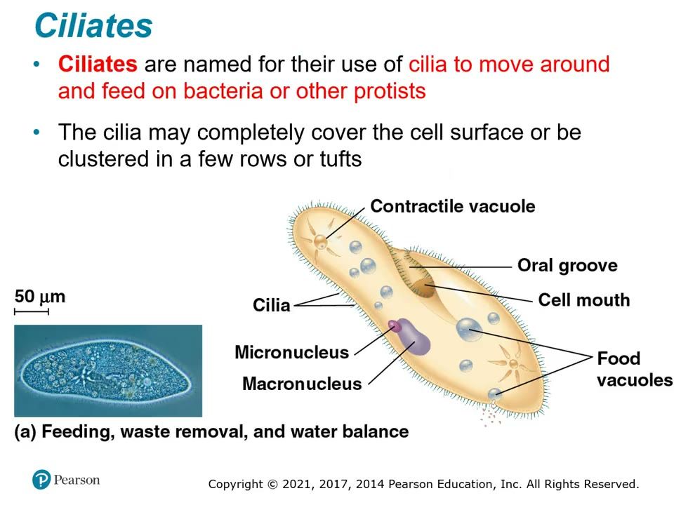
Slide 62. Paramecium, labeled
Ciliate: cilia over the whole surface, oral groove, cell mouth, food vacuoles, contractile vacuole, micronucleus and macronucleus.
How to draw it: Slipper-shaped cell fringed with cilia; oral groove on one side; large macronucleus, small micronucleus; label all.
▶ Play from 17:34Lecture 28.3, slide 020
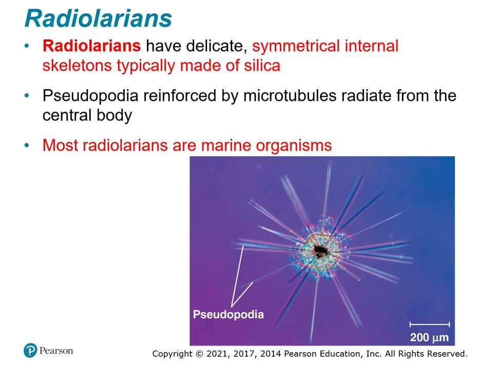
Slide 63. Radiolarian
Symmetrical internal skeleton usually of silica; thread-like pseudopodia reinforced by microtubules radiate out; mostly marine.▶ Play from 21:53Lecture 28.3, slide 030
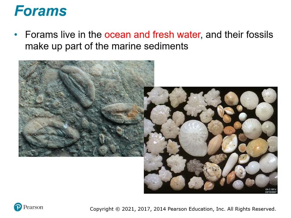
Slide 64. Forams
Porous calcium carbonate tests; ocean and fresh water; fossils form part of the marine sediments.▶ Play from 23:28Lecture 28.3, slide 032
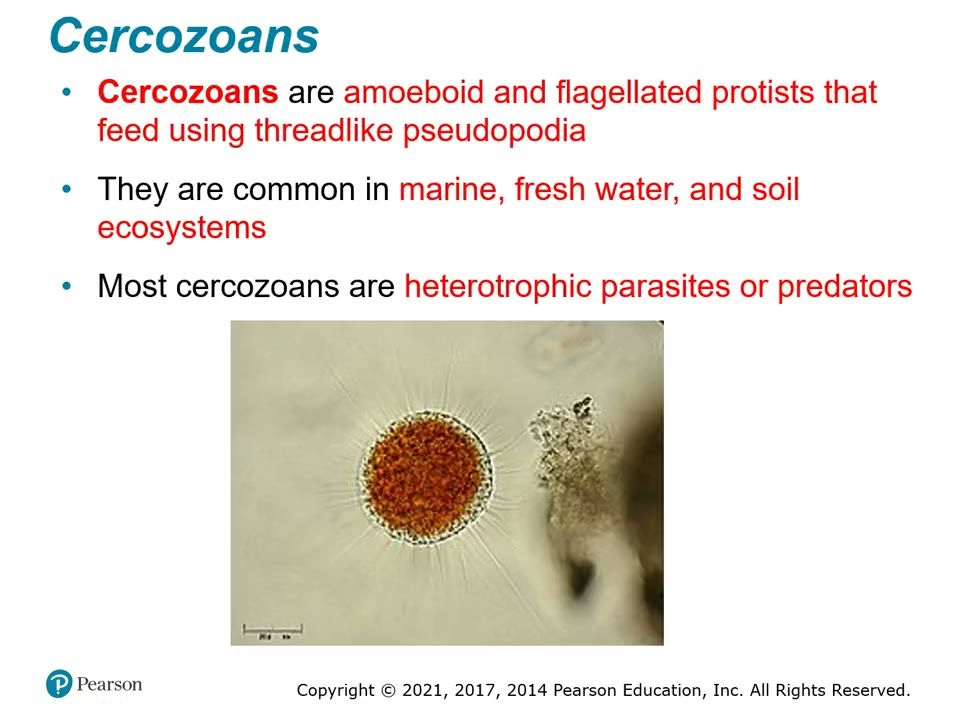
Slide 65. Cercozoan
Amoeboid and flagellated, thread-like pseudopodia; marine, freshwater and soil; heterotrophic parasites or predators.▶ Play from 24:22Lecture 28.3, slide 033
Be able to draw / explain
My notes
28.4

Red algae and green algae are the closest relatives of plants

Lecture video 28.4 (opens online)
Big idea
Archaeplastida contains red algae (a clade), green algae (a major group of chlorophytes and charophytes) and plants. Plastids arose when a heterotrophic protist took in a cyanobacterial endosymbiont; its photosynthetic descendants became the red and green algae, and plants descended from green algae. Red algae get their color from phycoerythrin; green algae have plant-like green chloroplasts.

Must know

Terms

Archaeplastida
Supergroup of red algae, green algae and plants.
phycoerythrin
Red accessory pigment of red algae that masks chlorophyll.
chlorophytes
Green algae clade, mostly freshwater (e.g. Volvox, Ulva, Caulerpa).
charophytes
Green algae clade most closely related to land plants.
paraphyletic
Includes an ancestor and some, but not all, of its descendants (green algae leave out plants).

Examples

What she stressed in lecture
Memory hooks

Slides

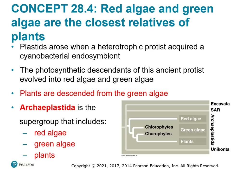
Slide 66. Archaeplastida tree
Plastids came from a cyanobacterial endosymbiont. Red algae, green algae (chlorophytes, charophytes) and plants; plants descended from green algae.▶ Play from 0:11Lecture 28.4, slide 002
Slide 67. Red algae
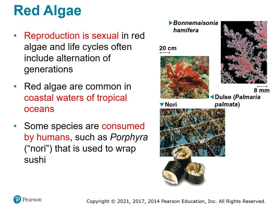
Sexual reproduction, often alternation of generations; common in warm coastal tropical waters; Porphyra (nori) wraps sushi; dulse.▶ Play from 2:55Lecture 28.4, slide 004
Slide 68. Green algae: three ways to get bigger
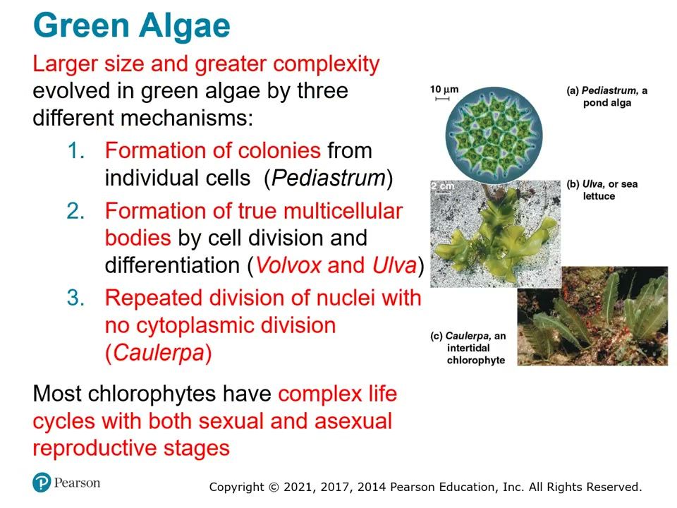
(1) Colonies of individual cells (Pediastrum); (2) true multicellular bodies (Volvox, Ulva); (3) repeated nuclear division with no cytoplasmic division (Caulerpa).▶ Play from 5:49Lecture 28.4, slide 007
Be able to draw / explain
My notes
28.5

Unikonts include protists that are closely related to fungi and animals

Lecture video 28.5 (opens online)
Big idea
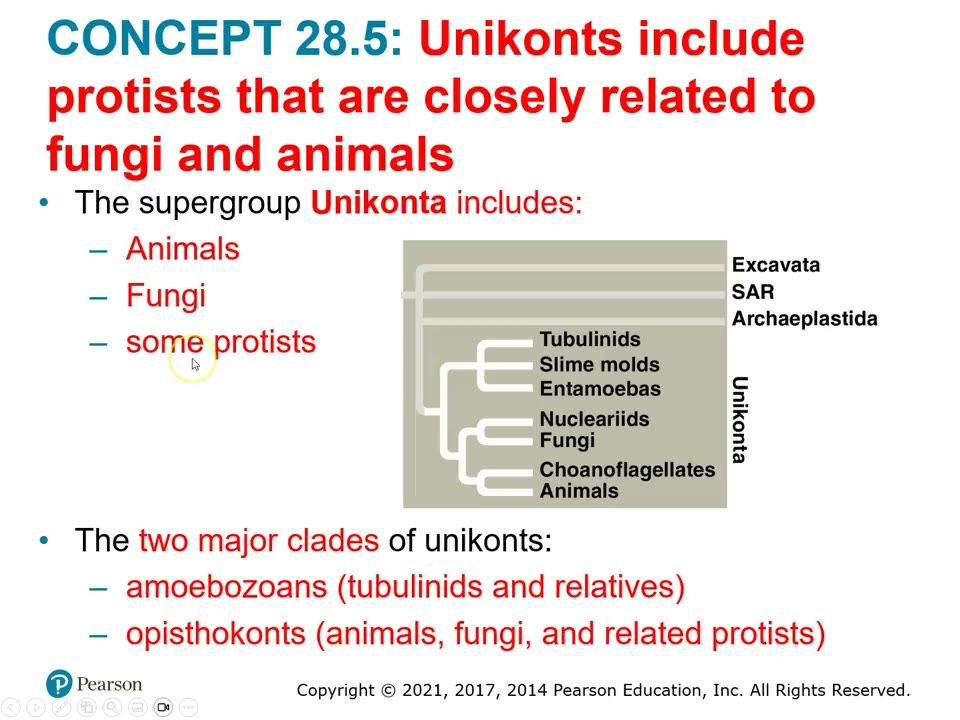
Unikonta contains animals, fungi and related protists in two major groups: Amoebozoans (amoebas with lobe- or tube-shaped pseudopodia: tubulinids, slime molds, entamoebas) and Opisthokonts (animals, fungi, choanoflagellates, nucleariids). One controversial hypothesis puts unikonts as the first eukaryotes to diverge.

Must know

Terms

Unikonta
Supergroup of amoebozoans and opisthokonts (animals, fungi and related protists).
Amoebozoa
Major group of amoebas with lobe- or tube-shaped pseudopodia.
Opisthokonta
Major group of animals, fungi and their protist relatives.
plasmodium (slime mold)
Single huge multinucleate (diploid) feeding mass of a plasmodial slime mold. Not the same as the malaria genus Plasmodium.
fruiting body
Stalked spore-producing structure slime molds form when conditions get bad.
detritus
Dead organic material, usually on the bottom of a lake or ocean.

Examples

What she stressed in lecture
Memory hooks

Slides

Slide 69. Unikonta tree
Two major groups: amoebozoans (tubulinids, slime molds, entamoebas) and opisthokonts (nucleariids, fungi, choanoflagellates, animals).▶ Play from 0:32Lecture 28.5, slide 004
Slide 70. Hypothesis: unikonts diverged first
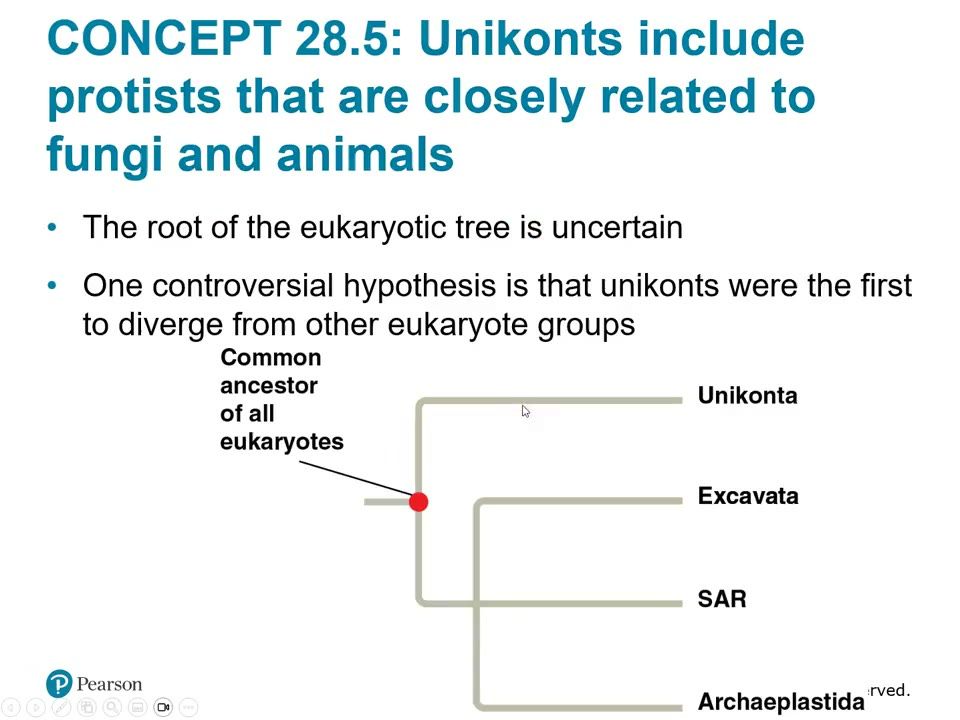
The root of the eukaryotic tree is uncertain; one controversial hypothesis puts unikonts as the first group to branch from the common ancestor of all eukaryotes.▶ Play from 1:35Lecture 28.5, slide 005
Slide 71. Plasmodial slime mold
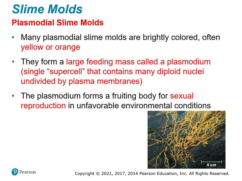
Bright yellow or orange plasmodium: one supercell with many diploid nuclei, undivided by membranes; forms fruiting bodies when conditions are poor.▶ Play from 4:53Lecture 28.5, slide 009
Slide 72. Cellular slime mold life cycle
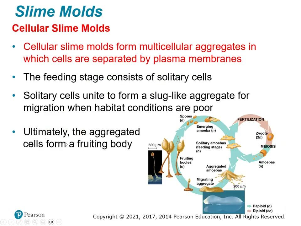
Solitary feeding cells aggregate into a slug-like mass (cells keep their membranes) that forms a fruiting body.▶ Play from 5:59Lecture 28.5, slide 010
Be able to draw / explain
My notes
28.6

Protists play key roles in ecological communities

Lecture video 28.6 (opens online)
Big idea
Protists live in aquatic and moist terrestrial habitats and act as symbionts (mutualists or parasites) or as producers. Photosynthetic protists and prokaryotes are the main producers in aquatic communities. Warming sea surfaces reduce the growth of photosynthetic protists, which can affect fisheries and the carbon cycle.

Must know

Terms

symbiont
An organism living in close association with another (mutualist or parasite).
producer
Organism that makes organic compounds from CO2 (photosynthesis).
consumer
Organism that eats producers directly or indirectly.

Examples

What she stressed in lecture
Memory hooks

Slides

Slide 73. Termite gut protist
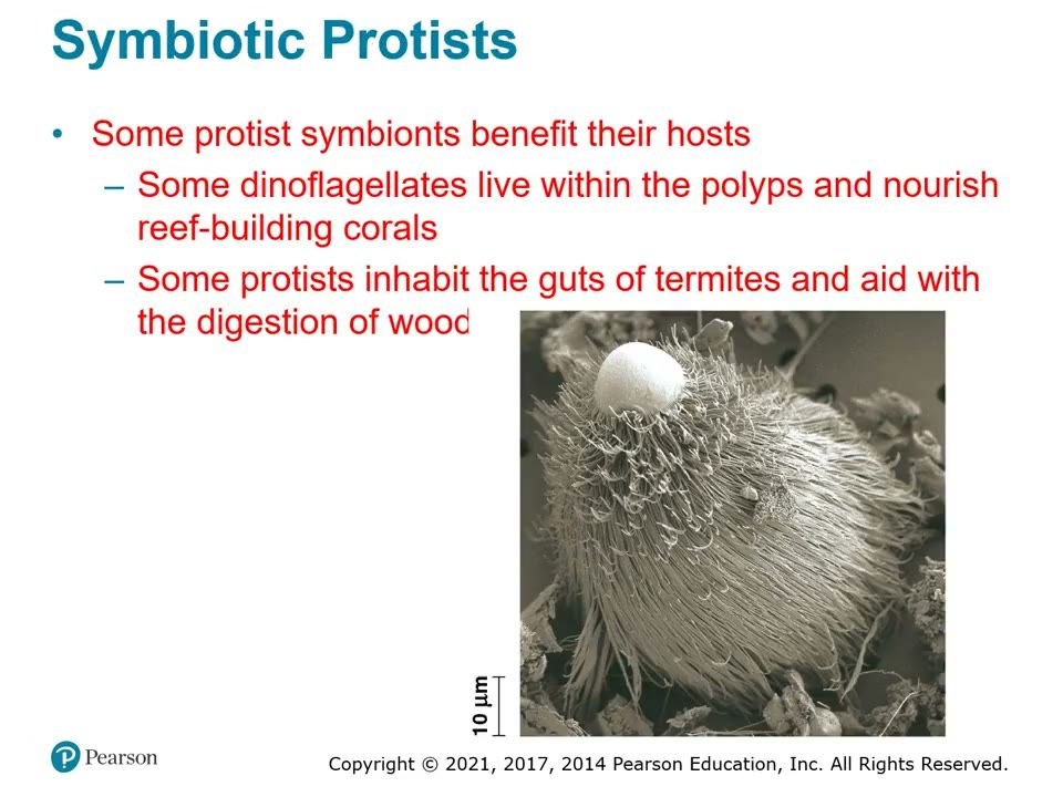
Mutualist that digests wood inside termites; dinoflagellates nourish reef-building corals.▶ Play from 0:50Lecture 28.6, slide 003
Slide 74. Sudden oak death
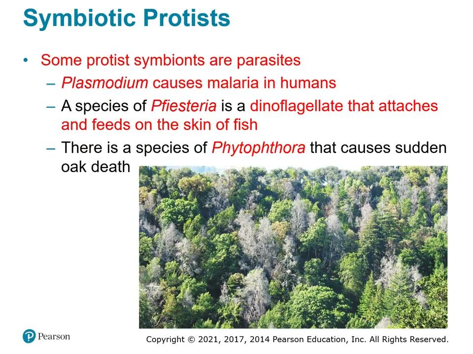
A species of Phytophthora kills oaks; Pfiesteria (dinoflagellate) feeds on fish skin; Plasmodium causes malaria.▶ Play from 1:50Lecture 28.6, slide 004
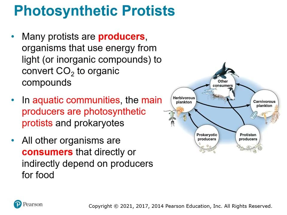
Slide 75. Producers in an aquatic food web
Photosynthetic protists and prokaryotes are the main producers; all other organisms are consumers.▶ Play from 2:55Lecture 28.6, slide 005
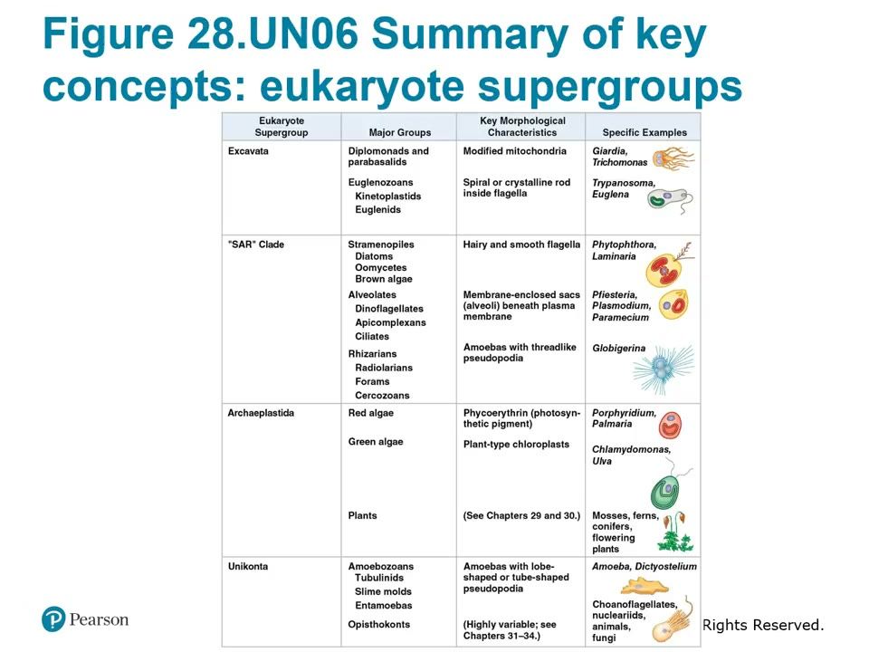
Slide 76. Figure 28.UN06 Summary of key concepts
Chapter summary table: supergroup, major groups, key morphological characteristics, specific examples. Suggested study sheet for Exam 2.▶ Play from 6:02Lecture 28.6, slide 007
My notes
Review

Compare tables, traps, essays

Bacteria vs Archaea vs Eukarya (focus on the differences)

CharacteristicBacteriaArchaeaEukarya
Cell typeProkaryoteProkaryoteEukaryote
Nuclear envelopeAbsentAbsentPresent
Membrane-enclosed organellesAbsentAbsentPresent
ChromosomeOne circular chromosome in nucleoidOne circular chromosome in nucleoidMultiple linear chromosomes in nucleus
Peptidoglycan in cell wallPresentAbsent (polysaccharides and proteins)Absent (cellulose in plants, chitin in fungi)
Membrane lipidsUnbranched hydrocarbonsSome branched hydrocarbonsUnbranched hydrocarbons
RNA polymeraseOne kindSeveral kindsSeveral kinds
Initiator amino acid for protein synthesisFormyl-methionineMethionineMethionine
PathogensAll known pathogenic prokaryotesNone knownSome
Examples from lectureProteobacteria, Chlamydias, Spirochetes, Cyanobacteria, Gram-positive bacteriaExtreme halophiles, extreme thermophiles, methanogens (Euryarchaeota, TACK, Lokiarchaeotes)Plants, fungi, animals, protists
Closest relativeBranched off firstEukaryaArchaea

Four nutritional modes

ModeEnergy sourceCarbon sourceExamples from lecture
PhotoautotrophLightCO2Cyanobacteria (Anabaena), some Proteobacteria, plants
ChemoautotrophInorganic chemicals (such as hydrogen sulfide)CO2Sulfur bacterium Thiomargarita, hydrothermal vent bacteria
PhotoheterotrophLightOrganic compoundsCertain prokaryotes only
ChemoheterotrophOrganic compoundsOrganic compoundsDecomposer prokaryotes, pathogens, spirochetes, fungi, animals

Gram-positive vs Gram-negative

FeatureGram-positiveGram-negative
PeptidoglycanThick layerThin layer
Outer membraneNoneOuter lipopolysaccharide membrane
Stain colorPurplePink/red
Antibiotic resistanceLess resistant (drugs reach peptidoglycan)More resistant (outer membrane blocks drugs)
EndotoxinsNo outer membrane lipopolysaccharideLipopolysaccharide released as endotoxin when cells die
Lecture groupsActinomycetes, Streptomyces, Staphylococcus, Bacillus, ClostridiumProteobacteria, Chlamydias, Spirochetes, Cyanobacteria
Oreo analogyOreo with one cookie removedWhole Oreo

Monophyletic vs paraphyletic vs polyphyletic

GroupIncludes most recent common ancestorIncludes all descendantsLecture example
Monophyletic (clade)YesYesEven-toed ungulates plus hippos plus cetaceans
ParaphyleticYesNoEven-toed ungulates plus hippos, leaving out cetaceans
PolyphyleticNoNot applicable (distantly related members)Cetaceans plus seals grouped by appearance

Transformation vs transduction vs conjugation

ProcessHow DNA movesKey detail
TransformationCell takes up foreign DNA from surroundingsCan turn a nonpathogenic cell pathogenic
TransductionBacteriophage carries genes between hostsUnintended result of phage replication
ConjugationDirect transfer between two joined cells via pilusOne direction only, donor to recipient; plasmids transferred

Orthologous vs paralogous genes

FeatureOrthologousParalogous
Origin of homologySpeciation eventGene duplication
Where foundIn different speciesCopies within one species
UseInferring phylogenyShow divergence within a genome (gene families)
ExampleCytochrome c in humans and dogsHuman olfactory receptor genes

Exotoxins vs endotoxins

FeatureExotoxinEndotoxin
What it isProtein secreted by bacteriaLipopolysaccharide of Gram-negative outer membrane
When releasedSecreted by living bacteriaOnly when bacteria die and walls break down
Needs bacteria presentNo, causes disease even after bacteria are goneReleased from bacterial cells
ExampleVibrio cholerae, choleraSalmonella, food poisoning

Oxygen relations

TypeOxygen use
Obligate aerobeRequires oxygen for cellular respiration
Obligate anaerobeCannot use oxygen; poisoned by it; anaerobic respiration
Facultative anaerobeUses oxygen if present; otherwise fermentation or anaerobic respiration

Major bacterial groups

GroupGramKey traitsNamed examples
ProteobacteriaNegativePhotoautotrophs, chemoautotrophs, heterotrophsNeisseria gonorrhoeae (gonorrhea), Vibrio cholerae (cholera), Helicobacter pylori (ulcers), Thiomargarita
ChlamydiasNegativeParasites of animal cells; walls lack peptidoglycanChlamydia trachomatis (nongonococcal urethritis)
SpirochetesNegativeHelical heterotrophs; internal filaments; free-living or pathogenicTreponema (syphilis), Borrelia (Lyme disease)
CyanobacteriaNegativePhotoautotrophs; solitary or filamentousAnabaena (heterocysts)
Gram-positive bacteriaPositiveVery diverse; actinomycetes form colonies, soil decomposers, pathogensStreptomyces (antibiotics), Staphylococcus aureus, Bacillus anthracis (anthrax), Clostridium botulinum (botulism)

Archaeal groups

GroupMembers / habitat
Extreme halophilesVery salty environments
Extreme thermophilesVery hot environments, over 100 degrees C
MethanogensObligate anaerobes making methane; under Greenland ice, swamps, marshes, guts of cattle, termites, herbivores
EuryarchaeotaExtreme halophiles, most methanogens, some extreme thermophiles
TACK supergroupThaumarchaeota, Aigarchaeota, Crenarchaeota (rest of extreme thermophiles), Korarchaeota
LokiarchaeotesClose to TACK; possible sister group of eukaryotes

History of classification schemes

SystemGroups
Two kingdomsPlants, animals (fungi and prokaryotes with plants)
Five kingdoms (by 1960s)Monera, Protista, Plantae, Fungi, Animalia
Three domains (current)Bacteria, Archaea, Eukarya (Plantae, Fungi, Animalia; Protista no longer a kingdom)
Traps (ch26-27)

Essay / short-answer prompts

Use this character table to build a phylogenetic tree and explain your reasoning. Taxa: lancelet, lamprey, bass, frog, turtle, leopard. Characters: vertebral column, hinged jaws, four limbs, amnion, hair (lancelet has none; lamprey has vertebral column only; bass adds hinged jaws; frog adds four limbs; turtle adds amnion; leopard adds hair). Identify the outgroup, the shared ancestral and shared derived characters, and the sister taxa.

Define monophyletic, paraphyletic, and polyphyletic groups. Draw one tree and circle an example of each. Give a real example of a paraphyletic and a polyphyletic group from lecture.

Distinguish homology from analogy. Explain why systematists must separate them before building a tree, how they tell them apart, and give examples of each from lecture and the videos.

Compare the cell walls of Gram-positive and Gram-negative bacteria. Explain how the Gram stain distinguishes them, why Gram-negative bacteria are more resistant to antibiotics, why peptidoglycan-targeting antibiotics do not harm humans, and how the Gram-negative wall relates to endotoxins.

Prokaryotes reproduce asexually by binary fission, yet their populations are highly diverse. Explain the three factors that produce this diversity and describe the three mechanisms of genetic recombination.

Describe the four nutritional modes of prokaryotes and give an example of each type of organism from lecture. Then explain oxygen relations and nitrogen fixation, including the role of heterocysts.

The four supergroups (Fig. 28.5)

SupergroupMajor groups / cladesDefining featureExamples
ExcavataDiplomonads, parabasalids, euglenozoans (kinetoplastids + euglenids)Cytoskeleton; some have an excavated feeding groove; modified mitochondria or unique flagellaGiardia, Trichomonas, Trypanosoma, Euglena
SARStramenopiles (diatoms, brown algae, oomycetes); Alveolates (dinoflagellates, apicomplexans, ciliates); Rhizarians (radiolarians, forams, cercozoans)Grouped by DNA similarityDiatoms, kelp, Phytophthora, red tide, Plasmodium, Paramecium, forams
ArchaeplastidaRed algae; green algae (chlorophytes, charophytes); plantsPlastids from a cyanobacterial endosymbiontNori (Porphyra), Pediastrum, Volvox, Ulva, Caulerpa
UnikontaAmoebozoans (tubulinids, slime molds, entamoebas); Opisthokonts (animals, fungi, choanoflagellates, nucleariids)Lobe/tube pseudopodia (amoebozoans); closest to fungi and animalsAmoeba proteus, slime molds, Entamoeba histolytica

Diseases and the protist that causes them

Disease / effectOrganismGroup
Intestinal infection (giardiasis)Giardia intestinalisExcavata: diplomonad
Sexually transmitted infectionTrichomonas vaginalisExcavata: parabasalid
Sleeping sickness; Chagas diseaseTrypanosomaExcavata: kinetoplastid
Potato late blightPhytophthora infestansSAR: oomycete
Sudden oak deathPhytophthoraSAR: oomycete
Red tide (toxic bloom)DinoflagellatesSAR: alveolate
Fish skin attacksPfiesteriaSAR: dinoflagellate
MalariaPlasmodiumSAR: apicomplexan
Amoebic dysenteryEntamoeba histolyticaUnikonta: entamoeba

Reduced mitochondria and special structures

StructureWho has itWhat it is
MitosomeDiplomonadsReduced mitochondrion, no electron transport chain
HydrogenosomeParabasalidsReduced mitochondrion, releases H2
KinetoplastKinetoplastidsOrganized DNA mass in the single mitochondrion
Crystalline/spiral rod in flagellumEuglenozoansDefining feature of the major group
AlveoliAlveolatesSacs under the plasma membrane
ApexApicomplexansOrganelle complex to penetrate host cells
Silica wall (2 parts)DiatomsGlass-like protective wall
Silica skeletonRadiolariansSymmetrical internal skeleton
Calcium carbonate testForamsPorous shell
Traps (ch28)

Essay / short-answer prompts

Describe the life cycle of Plasmodium. Name each stage, its host and ploidy, and say which part is sexual and which is asexual.

Name the four supergroups of eukaryotes and give the major groups or clades and one example in each.

Compare oomycetes and slime molds with true fungi. Why were they once classified as fungi, and why are they not fungi?